Southern Blot

Introduction

A Southern blot is a laboratory method used to detect specific DNA molecules among many other DNA molecules. The technique is named after its inventor, Edward Southern. As a laboratory procedure, Southern blots can be used to analyze an organism’s total DNA, also known as its genome, to identify a specific sequence of interest.

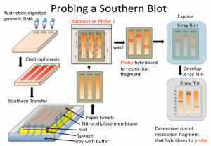
The first step in a Southern blot is to prepare the DNA mixture by breaking it into small fragments using a protein called a restriction enzyme. The mixture of DNA fragments is then separated by size using a technique called gel electrophoresis. After separation, the double-stranded DNA pieces denature or separate into single strands within the gel. The DNA is then transferred from the gel to a transfer membrane. Although this step is what gives the technique the name “southern transfer,” the term is generally used to describe the entire procedure.

Once the transfer is complete, the membrane carries all the bands originally on the gel. The membrane is then treated with a small piece of DNA or RNA called a probe, which has been designed to have a sequence that is complementary to a particular DNA sequence in the sample; this allows the probe to hybridize or bind to a specific DNA fragment on the membrane. Also, the probe has a label, which is usually a radioactive atom or a fluorescent dye. Thus, after hybridization, the probe allows the DNA fragment of interest to be detected among the many different DNA fragments on the membrane.

General procedure for drying

1. Sample homogenization involving DNA/RNA/protein purification that is performed after extraction from a variety of sources, such as cells or tissues.

2. Digest DNA with restriction enzymes into fragments, which is not necessary for RNA (northern blot).

3. Separation of the molecule of interest by membrane electrophoresis, generally on agarose gel for DNA fragments. In the case of RNA samples, they can be separated on an agarose gel in the presence of formaldehyde as a denaturing agent. This is necessary since formaldehyde limits the secondary structures of RNA molecules.

4. Transfer the molecules (DNA/RNA fragments) to a nitrocellulose membrane/nylon membrane from the gel.

5. Prehybridization (Blocking): Washing the nylon membrane with a prehybridization or blocking solution comprising salmon sperm DNA is required to block non-specific DNA interactions and also helps reduce background noise. Alternatively, there are some commercially available blocking buffers, such as PerfectHyb™ Plus buffer, in which salmon sperm DNA is not required for blocking purposes.

6. For probe preparation, a fresh 32P alpha-labelled dCTP-labeled DNA probe is prepared.

7. Hybridization or identification of the molecule that is achieved by incubating the blot with the specific labelled probe.

8. For detection of the probe and the DNA/RNA sequence of interest, the film is exposed to -80°C.

Beginning

Restriction endonucleases, which is enzymes, are used to break DNA into small fragments. These fragments are then separated by electrophoresis. The fragments obtained are then classified according to their size (kDa). Thus, the DNA fragments are transferred to blotting paper where they are incubated with probes. The probes used in Southern blotting can be very selective. They can selectively bind with a resolution of 1 in a million and the characteristics to bind to the intended target fragments.
Necessary materials reagents

  • The buffer used for electrophoresis is TAE or TBE.
  • The preferred agarose should be electrophoresis grade.
  • For DNA staining, ethidium bromide (0.5 µg ml–1, dissolved in H2O) is used. But there are other DNA staining dyes, such as SYBR green, that can replace ethidium bromide for safe handling.
  • 2X and 20X SSC (20X SSC composition includes 3.0 M NaCl and 0.3 M sodium citrate)
  • 6X DNA Loading Buffer consisting of 0.25% Bromophenol Blue, 0.25% Xylene Cyanol FF, and 30% Glycerol in water.
  • Suitable DNA markers of variable molecular weight, also called DNA ladders, are used as reference standards.
  • The prehybridization and hybridization mixture consists of 0.5% SDS, 6X SSC, 5X Denhardt’s solution, and 100 mg ml–1 sheared denatured salmon sperm DNA of yeast tRNA.
  • The widely used Denhardt solution for hybridization is composed of 0.02% Ficoll, 0.02% polyvinylpyrrolidone, and 0.02% bovine serum albumin (BSA)
  • Paraffin oil
  • Cellulose nitrate or nitrocellulose membrane filter with uniform porosity. (eg, Millipore 25 HAWP; nylon membranes used for blot protocols are available under various trade names from commercial vendors)
  • RNase A (20 pg ml–1 in 2X SSC) is required for specific cleavage.
  • Restriction enzyme and an appropriate buffer are used.
  • Radioactively labelled RNA as a probe for specific detection where autoradiography is performed.
  • For the detection of RNA labelled with labels such as 3H, 35S, 125I or 14C, 2,5-diphenyloxazole (PPO) in toluene at a concentration of 20% w/vol is required.

Team

1. Transfer of narrow strips of gel can be achieved using three pieces of glass or Plexiglas that are 5 cm × 20 cm in size with a thickness similar to the thickness of the gel.

2. There is a requirement for coarse, dry filter paper (four to five in number) or paper towels 10 cm × 18 cm in size.

3. The hybridization dish has a larger dimension (0.8 mm depth × 2 cm height × ~1 cm length) than the membrane used for hybridization, and the material used to develop the hybridization dish is Perspex (note: several alternative procedures for hybridization follow)

4. Four narrow pieces of Perspex that are similar in thickness to gel. Perspex length is sufficient to surround the gel at a distance of ~3 mm

5. A tray 20–50 mm (approx.) deep and 20 mm (approx.) in length and width larger than the gel

6. A sheet of glass long enough to fit in the pan and narrower to have a 10mm gap on each side

7. Several thick pieces of filter paper have a large area compared to the gel. The length of the filter paper is suitable to cover the glass sheet and can be immersed inside the tray.

8. A moistened piece of nitrocellulose membrane, which has a wider area that can cover the entire gel. The nitrocellulose membrane is placed on top of four strips of Perspex. Wetting of the nitrocellulose membrane is done using 2X SSC.

9. Paper towels that are stacked on top of each other.

10. Apparatus for straining gel.

11. A gel tank is necessary to perform electrophoresis.

12. Power supply is required for all device configurations.

Reagent Configuration

  • DNA: The entire procedure begins using DNA digested with enzymes of varying concentrations that will quantify the optimal concentration of DNA and specify the restriction enzyme to be used. Generally, an amount of 1 µg of DNA derived from clones (eg, from plasmid or bacteriophage clones) is sufficient for plasmids having low copy numbers. We need larger amounts to carry out the separation of complex DNA (eg genomic DNA). The advisable range to consider would be 5 to 10 µg.
  • The electrophoresis buffer used is TAE, which has a composition of 40 mM Tris, 20 mM acetic acid, 1 mM EDTA with a pH range of 7.4 to 8.2, which is normally prepared as a stock concentration of 20X or TBE composed of 89 mM Tris, 2.5 mM EDTA, 89 mM borate, normally made as a 10× stock)
  • TAE is recommended to be better when running gels for a shorter time interval and when recovery of DNA fragments from the gel is to be carried out.
  • TBE is considered to be a better buffer, especially when we have to run the gels for longer than 2 h.

Request

Southern blot is used in various applications. The main use of the Southern blot is to identify a specific DNA in a DNA sample. It is mainly used in the identification of viral infections and certain bacterial infections. In rDNA technology, the Southern blot technique is used to isolate a particular DNA. It is also useful in the study of genetic mutations and rearrangements, this property is used to diagnose neonatal diseases and genetic diseases. Due to the accuracy in DNA identification, this technique is used in phylogenetic studies, paternity and maternity analysis, forensic studies and personal identification.

Southern blotting can be applied to study the structure of a gene or to elucidate restriction enzyme maps. In particular, Southern blotting can be used to identify the methylated sites present for particular genes. This can be implemented by applying restriction nucleases such as MspI and HpaII, which can identify and cleave between identical sequences. The discovery of RFLPs by Southern blotting has aided in the mapping of several genomes that were crucial to map. In the field of immunology, clonal rearrangements of immunoglobulins, as well as T-cell receptor genes that play an important role in eliciting an immune response, can be analyzed by Southern blotting.

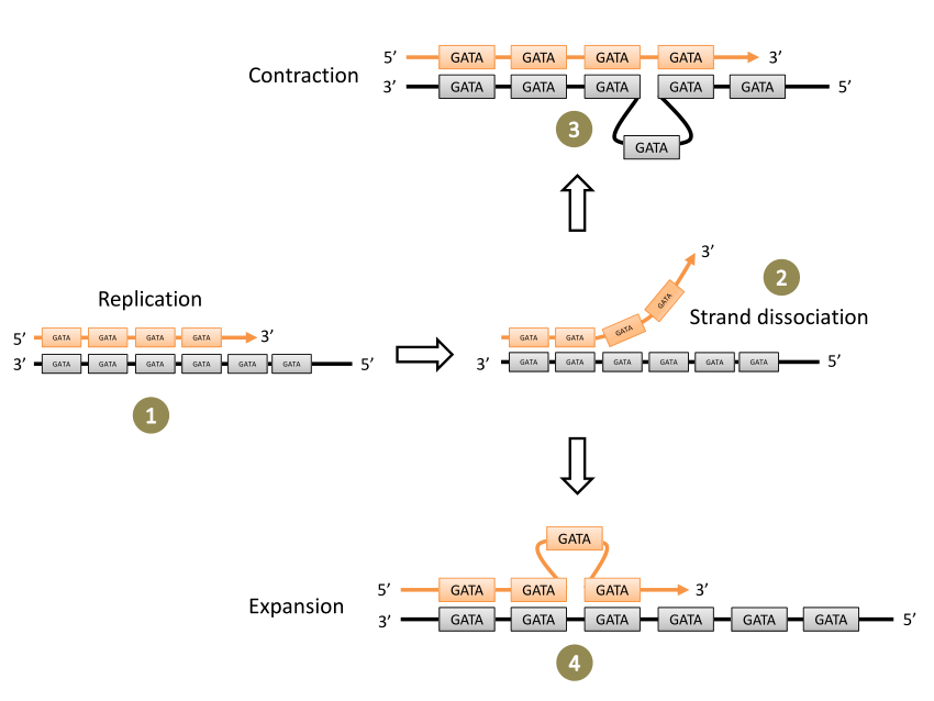
Slipped-strand mispairing

Abstract

Simple repetitive DNA sequences are a widespread and abundant feature of genomic DNA. The following features characterize such sequences: (1) they typically consist of a variety of 1-10 base repeat motifs, but may also include much larger repeats; (2) larger repeating units often include shorter ones within them; (3) long polypyrimidine and poly-CA tracts are often found; and (4) tandem arrangements of closely related motifs are often found. We propose that slipped strand mismatch events, together with unequal crossing over, can easily explain all of these features.

The frequent occurrence of long tandem repeats of particular motifs (polypyrimidine and poly-CA tracts) appears to be the result of non-random patterns of nucleotide substitution. We argue that the intrahelical process of slipped strand mismatching is much more likely to be the major factor in the initial expansion of short repeat motifs and that, after the initial expansion, simple tandem repeats may be predisposed to further expansion by unequal crossover or other interhelical events due to their propensity for mismatching.

Evidence is presented that single base repeats (the shortest possible motifs) are represented by longer series in mammalian introns than would be expected randomly, supporting the idea that SSM may be a ubiquitous force in the evolution of the eukaryotic genome. Thus, simple repetitive sequences may represent a natural ground state of DNA not selected for coding functions.

Key points

  • Altered gene expression is the result of SSM and, depending on where the increase or decrease in short repeat sequences occurs relative to the promoter, will be regulated at the level of transcription or translation. The result is an ON or OFF phase of a gene or genes.
  • SSM can result in an increase or decrease in the number of short repeat sequences. Short repeat sequences are from 1 to 7 nucleotides and can be homogeneous or heterogeneous repetitive DNA sequences.
  • Transcriptional regulation can occur if the repeats are located in the promoter region at the RNA polymerase binding site, -10 and -35 upstream of the gene(s).
  • SSM induces transcriptional regulation by changing short repeat sequences located outside the promoter. If there is a change in the short repeat sequence, it can affect the binding of a regulatory protein, such as an activator or a repressor.

Key terms

Slipped strand mismatch: a process that produces a mismatch of short repeat sequences between the parent strand and the daughter strand during DNA synthesis.

7.1D: Slipped-Strand Mispairing - Biology LibreTexts

Slipped strand mismatch (SSM) is a process that results in the mispairing of short repeated sequences between the mother and daughter strand during DNA synthesis. This RecA-independent mechanism can occur during DNA replication or DNA repair and can be on the leading or lagging strand and can result in an increase or decrease in the number of short repeat sequences. Short repeat sequences are from 1 to 7 nucleotides and can be homogeneous or heterogeneous repetitive DNA sequences.

Altered gene expression is the result of SSM and, depending on where the increase or decrease in short repeat sequences occurs relative to the promoter, will be regulated at the level of transcription or translation. The result is an ON or OFF phase of a gene or genes. Transcriptional regulation occurs in several ways. One possibility is if the repeats are located in the promoter region at the RNA polymerase binding site, -10 and -35, upstream of the gene(s). The opportunistic pathogen H. influenza has two divergently oriented promoters in the gene shift and hifB fimbriae.

Overlapping promoter regions have TA dinucleotide repeats at -10 and -35 sequences. Via SSM, the TA repeat region can undergo the addition or subtraction of TA dinucleotides, resulting in the reversible ON or OFF phase of hifA and hifB transcription. The second way that SSM induces transcriptional regulation is by changing short repeat sequences located outside of the promoter. If there is a change in the short repeat sequence, it can affect the binding of a regulatory protein, such as an activator or a repressor. It can also give rise to differences in the post-transcriptional stability of the mRNA.

Restriction Fragment Length Polymorphisms (RFLPs)

Abstract

Restriction fragment length polymorphisms, or RFLPs, are differences between individuals in the lengths of DNA fragments cut by enzymes. Restriction enzymes are proteins that cut DNA at short, specific sequences called restriction sites. After cutting a segment of DNA with restriction enzymes, researchers can examine the fragments using a laboratory method called gel electrophoresis, which separates DNA fragments based on their size.

If two individuals have differences in their DNA sequences at particular restriction sites, the restriction enzymes will cut their DNA into fragments of different lengths. There may also be differences in the number of DNA fragments observed between two or more individuals. Gentaur RFLP analysis can be used as a form of genetic testing to see if an individual carries a mutant gene for a disease that runs in their family.

Beginning

Restriction endonucleases are enzymes that cut long DNA into short pieces. Each restriction endonuclease targets different nucleotide sequences in a DNA strand and therefore cuts at different sites. The distance between the cleavage sites of a given restriction endonuclease differs between individuals. Therefore, the length of the DNA fragments produced by a restriction endonuclease will differ between individual organisms and species.

How does it work?

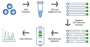
The RFLP is carried out through a series of steps that are briefly described below:

  • Extraction of DNA

To begin with, DNA is extracted from blood, saliva, or other samples and purified.

  • DNA fragmentation

The purified DNA is digested using restriction endonucleases. The recognition sites of these enzymes are generally 4 to 6 base pairs in length. The shorter the recognized sequence, the greater the number of fragments generated from digestion.

For example, if there is a short GAGC sequence that occurs repeatedly in a DNA sample. The restriction endonuclease that recognizes the GAGC sequence cuts the DNA at each repeat of the GAGC pattern. If one sample repeats the GAGC sequence 4 times while another sample repeats it 2 times, the length of the fragments generated by the enzyme for the two samples will be different.

  • Gel electrophoresis

Restriction fragments produced during DNA fragmentation are analyzed by gel electrophoresis. The fragments are negatively charged and can be easily separated by electrophoresis, which separates molecules based on their size and charge. The fragmented DNA samples are placed in the chamber containing the electrophoretic gel and two electrodes. When an electric field is applied, the fragments migrate towards the positive electrode. Smaller fragments move faster through the gel, leaving larger ones behind, and therefore the DNA samples separate into distinct bands on the gel.

  • Band Visualization

The gel is treated with luminescent dyes to make the DNA bands visible.

RFLP Applications

RFLP has been used for various genetic analysis applications since its invention.

Some of these key RFLP applications are listed below:

  • Determine the status of genetic diseases such as Cystic Fibrosis in an individual.
  • To determine or confirm the origin of a DNA sample, such as in paternity tests or criminal investigations.
  • In genetic mapping to determine recombination rates showing the genetic distance between loci.
  • To identify a carrier of a disease-causing mutation in a family.

RFLP Disadvantages

1. Since its invention, RFLP has been a widely used genome analysis technique in forensic science, medicine, and genetic studies. However, it has become almost obsolete with the advent of relatively simple and less expensive DNA profiling technologies, such as polymerase chain reaction (PCR).

2. The RFLP procedure requires numerous steps and takes weeks to obtain results, while techniques such as PCR can amplify target DNA sequences in a few hours.

3. In addition, RFLP requires a large DNA sample, the isolation of which can be a laborious and time-consuming process. In contrast, PCR can amplify minute amounts of DNA in a matter of hours.

4. Due to numerous reasons like these, the PCR technique has largely replaced RFLP in most applications that require DNA sequencings, such as paternity testing or forensic sample analysis.

5. Furthermore, the identification of single nucleotide polymorphisms in the Human Genome Project has nearly replaced the need for RFLP in disease state analysis.

Diagnostic Accuracy of a Direct Panfungal Polymerase Chain Reaction Assay Performed on Stained Cytology Slides

Diagnostic Accuracy of a Direct Panfungal Polymerase Chain Reaction Assay Performed on Stained Cytology Slides

Molecular strategies are more and more being utilized to stained cytology slides for the analysis of neoplastic and infectious ailments. Such strategies for the identification of fungi from stained cytology slides haven’t but been evaluated. This research aimed to evaluate the diagnostic accuracy of direct (with out nucleic acid isolation) panfungal polymerase chain response (PCR) adopted by sequencing for identification of fungi and oomycetes on stained cytology slides from canines, cats, horses, and different species. Thirty-six circumstances had been recognized with cytologically identifiable fungi/oomycetes and concurrent identification through fungal tradition or immunoassay.

Twenty-nine controls had been recognized with no cytologically or histologically seen organisms and a concurrent adverse fungal tradition. Direct PCR concentrating on the inner transcribed spacer area adopted by sequencing was carried out on one cytology slide from every case and management, and the sensitivity and specificity of the assay had been calculated. The sensitivity of the panfungal PCR assay carried out on stained cytology slides was 67% total, 73% excluding circumstances with oomycetes, and 86% when contemplating solely slides with plentiful fungi. The specificity was 62%, which was attributed to amplification of fungal DNA from management slides with no seen fungus and adverse tradition outcomes.

Workplace-collected blood spots deposited on filter paper had been analysed with multiplexed affinity-based protein assays and located to be appropriate for proteomics evaluation. The protein extension assay (PEA) was used to characterize 92 proteins utilizing 1.2 mm punches in repeated samples collected from 20 employees. Overall, 97.8% of the samples and 91.3% of the analysed proteins handed high quality management. Both inside and between spot correlations utilizing six replicates from the identical particular person had been above 0.99, suggesting that comparable ranges are obtained from a number of punches from the identical spot and from consecutive spots.

Protein ranges from dried blood and moist serum from the identical people had been in contrast and the bulk of the analysed proteins had been discovered to be considerably correlated. These outcomes open up for simplified pattern assortment of blood in discipline circumstances for proteomic evaluation, but additionally spotlight that not all proteins might be robustly measured from dried complete blood. Direct panfungal PCR is succesful of offering genus- or species-level identification of fungi from stained cytology slides. Given the potential of panfungal PCR to amplify contaminant fungal DNA, this assay needs to be carried out on slides with seen fungi and interpreted along side morphologic evaluation by a medical pathologist.

Paper-based loop-mediated isothermal amplification and lateral stream (LAMP-LF) assay for identification of tissues of cattle origin

The current research was made with the aims of growth and standardization of cattle particular paper-based loop-mediated isothermal amplification cum lateral stream assay (LAMP-LFA), as a Point-of-care check (POCT) for identification of tissue of cattle origin. The parts of standardized LAMP response using cattle particular primer units had been lyophilized over paper buttons, recognized finest because the provider of LAMP reagents. Based on possible LAMP amplicon, a pair of probes was designed, tagged and its hybridization with the amplified product of paper LAMP response was optimized. The parts of lateral stream assay for detection of probe hybridized LAMP merchandise had been standardized. Analysis of profitable amplification was made by utilizing HNB dye, LAMP-LFA strip, and likewise by the standard ladder-like sample on gel electrophoresis.

The assay was discovered extremely particular for cattle with an analytical sensitivity of 0.1 pg of absolute DNA. Laboratory validation carried out on samples from completely different people of cattle, coded samples, binary meat admixture, and heat-processed cattle tissues substantiated the accuracy of the assay. Comparison with pre-standardized species-specific PCR assay taken as gold requirements revealed 100% conformity. The discipline utility of the developed assay was additional established by its compatibility with the business package eliminating the prolonged DNA extraction step and storage stability of LAMP reagent provider buttons for Four months below refrigeration. Thus, the developed assay succesful of the end result inside Three h in resource-limited settings can be utilized as POCT for identification of tissue of cattle origin.

 Diagnostic Accuracy of a Direct Panfungal Polymerase Chain Reaction Assay Performed on Stained Cytology Slides

High-Speed Lens-Free Holographic Sensing of Protein Molecules Using Quantitative Agglutination Assays

Accurate, cost-effective, easy-to-use, and point-of-care sensors for protein biomarker ranges are necessary for illness diagnostics. A cheap and compact readout method that has been used for a number of diagnostic functions is lens-free holographic microscopy, which offers an ultralarge discipline of view and submicron decision when it’s coupled with pixel super-resolution strategies. Despite its potential as a diagnostic method, lens-free microscopy has not beforehand been utilized to quantitative protein molecule sensing in answer, which might simplify sensing protocols and finally allow measurements of binding kinetics in physiological circumstances.

Here, we sense interferon-γ (an immune system biomarker) and NeutrAvidin molecules in answer by combining lens-free microscopy with a one-step bead-based agglutination assay, enabled by a customized high-speed light-emitting diode (LED) array and automatic picture processing routines. We name this a quantitative large-area binding (QLAB) sensor.

[Linking template=”default” type=”products” search=”Red Active Caspase-9 Staining Kit 25 assays” header=”1″ limit=”149″ start=”2″ showCatalogNumber=”true” showSize=”true” showSupplier=”true” showPrice=”true” showDescription=”true” showAdditionalInformation=”true” showImage=”true” showSchemaMarkup=”true” imageWidth=”” imageHeight=””]

The high-speed gentle supply offers, for the primary time, pixel super-resolved imaging of >104 2 μm beads in answer present process Brownian movement, with out vital movement blur. The automated picture processing routines allow the counting of particular person beads and clusters, offering a quantitative sensor readout that relies upon on each bead and analyte concentrations. Fits to the chemical binding idea are offered. For NeutrAvidin, we discover a restrict of detection (LOD) of <27 ng/mL (450 pM) and a dynamic vary of 2-Four orders of magnitude. For mouse interferon-γ, the LOD is <Three ng/mL (200 pM) and the dynamic vary is at the very least Four orders of magnitude. The QLAB sensor holds promise for point-of-care functions in low-resource communities and the place protocol simplicity is necessary.

Lysosome Targeting RedGreen-assay: Selective Autophagy Sensing Assay for Mammalian Cells

Lysosome Targeting RedGreen-assay: Selective Autophagy Sensing Assay for Mammalian Cells

The means of autophagy is a necessary mobile mechanism, required to take care of basic cell well being by the elimination of dysfunctional organelles, such because the ER, peroxisomes and mitochondria, in addition to protein aggregates, and micro organism. Autophagy is an especially dynamic course of, and instruments are continually being developed to check the varied steps of this course of. This protocol particulars a technique to check the top steps of autophagy-lysosomal fusion and the formation of the autolysosome. Many strategies have been used to check the varied steps of the autophagy course of.

Here we describe the RedGreen-assay (RG-assay), an immunofluorescence-based method used to visualise the concentrating on of substrates to the autolysosome in reside cells. This method takes benefit of the low lysosomal pH and over-expression of a tandem GFP-mCherry tagged protein focused to an organelle of curiosity. While within the impartial cytosol or autophagosome, each GFP and RFP will fluoresce. However, inside the autolysosome, the GFP sign is quenched as a result of low pH surroundings and the RFP emission sign will predominate. This method is quickly quantifiable and amenable to excessive throughput experiments. Additionally, by tagging the GFP-RFP tandem fluorescent protein with organelle particular concentrating on sequences, it may be used to measure a variety of substrates of autophagy.

Quantifying the ratio of alternatively spliced mRNA variants of genes with identified various splicing variants is extremely related for many purposes. Herein, we describe the validation of a quantitative PCR design for the simplified quantification of identified mRNA splice variants. The assay makes use of a single-common primer pair, twin probe design for the willpower of splicing variants in a single nicely configuration. We used murine XBP-1 splicing variants, XBP-1S and XBP-1U, to validate and show the efficiency traits of this method. Using artificial XBP-1S and XBP-1U cDNA in addition to cDNA synthesized from mouse beta-cell line MIN6, we established the efficiency parameters and dynamic vary of the assay. Reliable quantification of each variants at various focus gradients was proven.

Platelet Isolation and Activation Assays

Platelets regulate hemostasis and are the important thing determinants of pathogenic thrombosis following atherosclerotic plaque rupture. Platelets flow into in an inactive state, however develop into activated in response to wreck to the endothelium, which exposes thrombogenic materials resembling collagen to the blood circulate. Activation leads to plenty of responses, together with secretion of soluble bioactive molecules through the discharge of alpha and dense granules, activation of membrane adhesion receptors, launch of microparticles, and externalization of phosphatidylserine.

These processes facilitate agency adhesion to websites of damage and the recruitment and activation of different platelets and leukocytes, leading to aggregation and thrombus formation. Platelet activation drives the hemostatic response, and likewise contributes to pathogenic thrombus formation. Thus, quantification of platelet-associated responses is vital to many pathophysiologically related processes. Here we describe protocols for isolating, counting, and activating platelets, and for the fast quantification of cell floor proteins utilizing circulate cytometry.

Extracellular vesicles (EVs) are produced by all domains of life together with Bacteria, Archaea and Eukarya. EVs are important for mobile physiology and comprise assorted cargo: virulence components, cell wall transforming enzymes, extracellular matrix parts and even nucleic acids and metabolites. While varied protocols for isolating EVs have been established for mammalian cells, the sphere is actively growing instruments to check EVs in different organisms.

In this protocol we describe our strategies to carry out density gradient purification of EVs in bacterial cells, permitting for separation of EV subpopulations, adopted by safety assays for EV cargo characterization. Furthermore, we devised a protocol which includes a fluorescent conjugate of fatty acids into EVs, the primary to permit reside-cell EV monitoring to look at launch of EVs, together with throughout an infection of mammalian cells by pathogenic micro organism. These protocols are highly effective instruments for EV researchers as they allow the statement of EV launch and the research of the mechanisms of their formation and launch.

 Lysosome Targeting RedGreen-assay: Selective Autophagy Sensing Assay for Mammalian Cells

Ex vivo Drosophila Wing Imaginal Disc Culture and Furin Inhibitor Assay

Furin is an evolutionarily conserved proprotein convertase (PC) household enzyme with a broad vary of substrates which might be important for developmental, homeostatic, and illness pathways. Classical genetic approaches and in vitro biochemical or cell organic assays recognized that precursor types of most development issue household proteins are processed by Furin. To quantitatively assess the potential function of Furin in cleaving and modulating intercellular dispersion of a Drosophila signaling protein, we developed a easy assay by combining genetics, ex vivo organ tradition, pharmacological remedy, and imaging analyses.
[Linking template=”default” type=”products” search=”Caspase-9 Colorimetric Assay Kit – 25 assays” header=”3″ limit=”154″ start=”2″ showCatalogNumber=”true” showSize=”true” showSupplier=”true” showPrice=”true” showDescription=”true” showAdditionalInformation=”true” showImage=”true” showSchemaMarkup=”true” imageWidth=”” imageHeight=””]
The protocol herein describes easy methods to ex vivo tradition Drosophila wing imaginal discs expressing a fluorescently tagged Drosophila Fibroblast Growth Factor (FGF, Branchless/Bnl) over an extended time period within the presence of Furin inhibitors and monitor the cleavage and intercellular dispersion of the truncated Bnl components utilizing microscopy. Although the assay described right here is for assessing the impact of Furin inhibition on Bnl cleavage within the Drosophila larval wing imaginal disc, the precept and methodology can simply be adopted for every other alerts, tissue techniques, or organisms. This technique and protocol present an assay for analyzing Furin exercise on a particular substrate by straight visualizing the spatiotemporal distribution of its truncated components in an ex vivo-cultured organ.

A Highly Sensitive, Reproducible Assay for Determining 4-hydroxynonenal Protein Adducts in Biological Material

A Highly Sensitive, Reproducible Assay for Determining 4-hydroxynonenal Protein Adducts in Biological Material

Oxidative stress is related to quite a few illnesses, and markers of oxidative stress in organic materials have gotten a mainstay of each experimental and scientific/epidemiological analysis. Lipid peroxidation is a serious type of oxidative stress, however resulting from their speedy degradation and instability, lipid peroxides are notoriously tough to measure, significantly in organic specimens the place their manufacturing and removing are constantly occuring. Thus, a generally used surrogate marker of lipid peroxidation is protein adducts of 4-Hydroxynonenal (HNE), an α, β-unsaturated hydroxyalkenal (i.e., a reactive aldehyde) fashioned through degradation of oxidized polyunsaturated fatty acids (PUFAs).

HNE adducts will be measured through commercially-available immunosorbent assays, however these have their limitations resulting from extreme prices, and reproducibility amongst laboratories is difficult resulting from variability in assay sensitivity, process, and reagents. Here we current a reproducible, facile, and economically conservative protocol for quantifying HNE protein adducts. The key to this protocol is to generate HNE-adduct requirements by incubating bovine serum albumin (BSA) with HNE. These requirements are then adsorbed to immunsorbent plastic in a multi-well plate format alongside organic samples.

An enzyme-linked immunosorbent assay (ELISA) is then carried out on the multi-well plate utilizing commercially-available major and secondary antibodies, and a peroxide-based fluorescent growing reagent. This protocol is very delicate and provides benefits to business sources in that it permits for reproducible, high-throughput quantitation of HNE adducts in a lot of samples. As such, it might be helpful as a biomarker of power oxidative stress for experimental and scientific research.

Understanding the molecular mechanism governing the higher-order regulation of actin dynamics requires chemically-defined and quantitative assays. Recently, the membrane reworking giant GTPase, dynamin, has been recognized as a brand new actin cross-linking molecule. Dynamin regulates actin cytoskeleton via binding to, self-assembling round, and aligning them into actin bundles. Here we make the most of dynamin for example and current a easy protocol to investigate the actin bundling exercise in vitro. This protocol particulars the strategy for F-actin reconstitution in addition to quantitative and qualitative analyses for actin bundling exercise of dynamins. Measurement of the actin bundling exercise of different actin-binding proteins might also be utilized to this protocol with acceptable changes relying on the protein of curiosity.

Genetic identification of medicinally used Salacia species by nrDNA ITS sequences and a PCR-RFLP assay for authentication of Salacia-related well being meals

The roots and stems of a number of Salacia species have been used as conventional medicines, particularly in Ayurvedic medical system for the therapy of diabetes, rheumatism, gonorrhea, amenorrhea, pores and skin illnesses, and so forth. Due to reported proof supporting Salacia’s useful results in early-stage diabetes and different lifestyle-related illnesses, Salacia-based dietary dietary supplements and well being meals have been gaining reputation in Japan and different nations in current years. However, because of the morphological similarities between Salacia crops, significantly in the medicinally used elements (roots and stems), the authentication of the botanical identities of Salacia-derived merchandise is difficult. This research goals to develop a genetic strategy to authenticate the medicinally used Salacia species and to find out the botanical sources of the commercially obtainable Salacia-derived merchandise.
 The sequences of nuclear DNA inside transcribed spacer (ITS) and chloroplast trnK-rps16 area had been decided and in contrast between 10 plant specimens from three medicinally used Salacia species in addition to 48 samples of business crude medicine. Moreover, a PCR-restriction fragment size polymorphism (RFLP) assay was developed for speedy identification primarily based on the ITS sequences. The plant specimens from the three medicinally used Salacia species confirmed three foremost kinds of sequences in each ITS (sorts I, II, III) and trnK-rps16 (i, ii, iii) areas.
Combined the sequences of ITS and trnK-rps16 areas, S. reticulata and S. oblonga had sort I-i and kind III-iii or comparable sequences, respectively. S. chinensis had sort II-ii or II(536M)-i sequences. Forty-eight samples of business crude medicine had been recognized primarily based on ITS and trnK-rps16 DNA barcode. A handy PCR-RFLP assay utilizing Cac8I restriction enzyme was established and utilized to determine the botanical sources of well being meals merchandise bought from on-line retailers. All the twelve samples had been recognized as S. chinensis.
The nrDNA ITS sequences offered helpful info to authenticate Salacia species and to elucidate the phylogenetic relationship inside the Salacia genus. Genetic identification outcomes revealed that S. chinensis and S. reticulate are the most important sources of commercially obtainable Salacia-products. Based on the ITS sequences, a handy PCR-RFLP assay was established for the identification of the medicinally used Salacia species in addition to their derived well being meals merchandise.
 A Highly Sensitive, Reproducible Assay for Determining 4-hydroxynonenal Protein Adducts in Biological Material

The Ring-Pull Assay for Mechanical Properties of Fibrous Soft Tissues – An Analysis of the Uniaxial Approximation and a Correction for Nonlinear Thick-Walled Tissues

The ring-pull check, the place a hoop of tissue is hooked through two pins and stretched, is a well-liked biomechanical check, particularly for small arteries. Although handy and dependable, the ring check produces inhomogeneous pressure, making willpower of fabric parameters non-trivial. To decide correction elements between ring-pull-estimated and true tissue properties.  A finite-element mannequin of ring pulling was constructed for a pattern with nonlinear, anisotropic mechanical conduct typical of arteries. The pin power and pattern cross-section had been used to compute an obvious modulus at small and huge pressure, which had been in comparison with the desired properties. The ensuing corrections had been validated with experiments on porcine and ovine arteries.
[Linking template=”default” type=”products” search=”Alkaline Phosphatase Activity Fluorometric Assay Kit 500 assays” header=”2″ limit=”144″ start=”3″ showCatalogNumber=”true” showSize=”true” showSupplier=”true” showPrice=”true” showDescription=”true” showAdditionalInformation=”true” showImage=”true” showSchemaMarkup=”true” imageWidth=”” imageHeight=””]
The correction was additional utilized to experiments on mouse aortic rings to find out materials and failure properties. Calculating pressure primarily based on centerline stretch somewhat than inner-wall or outer-wall stretch afforded higher estimation of tissue properties. Additional correction elements had been developed primarily based on ring wall thickness H, centerline ring radius R c , and pin radius a. The corrected estimates for tissue properties had been in good settlement with uniaxial stretch experiments.

Gentamicin Protection Assay to Determine the Number of Intracellular Bacteria during Infection of Human TC7 Intestinal Epithelial Cells by Shigella flexneri

Gentamicin Protection Assay to Determine the Number of Intracellular Bacteria during Infection of Human TC7 Intestinal Epithelial Cells by Shigella flexneri

Shigella flexneri is an intracellular bacterial pathogen that positive aspects entry to the intestine epithelium utilizing a specialised Type III Secretion System (T3SS). Various determinants mediating this invasive an infection have been experimentally verified utilizing the classical gentamicin safety assay introduced right here. In this assay epithelial cell strains are contaminated by micro organism in vitro and the extracellular micro organism are killed by gentamicin. The internalized micro organism, that are shielded from the bactericidal motion of gentamicin, are recovered by lysing the epithelial cells and enumerated by figuring out the colonies fashioned on strong medium.

Various methods primarily based on gentle microscopy, reminiscent of immunofluorescence and micro organism expressing fluorescent proteins, are additionally used for finding out intracellular micro organism. However, these methods should not solely labor intensive and require refined tools, however principally are additionally not quantitative. Despite being a straightforward quantitative technique to research invasiveness of micro organism, the gentamicin safety assay can’t distinguish between the survival and multiplication of the internalized micro organism over longer incubation durations.

To alleviate the problems created by multiplication and dissemination of internalized micro organism, complementary assays like plaque formation assays are required. This protocol presents a straightforward and cost-effective technique to decide the invasiveness and the capability to set up an an infection of Shigella below totally different circumstances. Automatic and visible interpretation of outcome bands have been additionally in contrast for the immunochromatography-based BinaxNOW and ImmuView UATs.

Urinary antigen exams (UATs) are sometimes used to diagnose Legionnaires’ illness as they’re speedy and simple to carry out on readily obtainable urine samples with out the want for specialised abilities in contrast to standard strategies. Recently developed automated readers for UATs could present goal outcomes interpretation, particularly in instances of weak outcome bands. Using 53 outlined affected person urine samples, we evaluated the efficiency of the BinaxNOW Legionella Antigen Card (Abbott), ImmuView S. pneumoniae and Legionella (SSI Diagnostica), STANDARD F Legionella Ag FIA (SD Biosensor), and Sofia Legionella FIA (Quidel) concurrently with their respective automated readers.

SARS-CoV-2 lateral circulation assays for attainable use in nationwide covid-19 seroprevalence surveys (React 2): diagnostic accuracy research

ensitivity analyses have been carried out on sera saved from 320 earlier contributors in the React 2 programme with confirmed earlier extreme acute respiratory syndrome coronavirus 2 (SARS-CoV-2) an infection. Specificity analyses have been carried out on 1000 prepandemic serum samples. 100 new contributors with confirmed earlier SARS-CoV-2 an infection attended research clinics for finger prick testing.  Laboratory sensitivity and specificity analyses have been carried out for seven LFIAs on a minimal of 200 serum samples from contributors with confirmed SARS-CoV-2 an infection and 500 prepandemic serum samples, respectively.
Three LFIAs have been discovered to have a laboratory sensitivity superior to the finger prick sensitivity of the LFIA presently utilized in React 2 seroprevalence research (84%). These LFIAs have been then additional evaluated by way of finger prick testing on contributors with confirmed earlier SARS-CoV-2 an infection: two LFIAs (Surescreen, Panbio) have been evaluated in clinics in June-July 2020 and the third LFIA (AbC-19) in September 2020. A spike protein enzyme linked immunoassay and hybrid double antigen binding assay have been used as laboratory reference requirements.
The accuracy of LFIAs in detecting immunoglobulin G (IgG) antibodies to SARS-CoV-2 in contrast with two reference requirements. The sensitivity and specificity of seven new LFIAs that have been analysed utilizing sera diverse from 69% to 100%, and from 98.6% to 100%, respectively (in contrast with the two reference requirements). Sensitivity on finger prick testing was 77% (95% confidence interval 61.4% to 88.2%) for Panbio, 86% (72.7% to 94.8%) for Surescreen, and 69% (53.8% to 81.3%) for AbC-19 in contrast with the reference requirements. Sensitivity for sera from matched medical samples carried out on AbC-19 was considerably larger with serum than finger prick at 92% (80.0% to 97.7%, P=0.01). Antibody titres diverse significantly amongst cohorts. The numbers of constructive samples recognized by finger prick in the lowest antibody titre quarter diverse amongst LFIAs.
Gentamicin Protection Assay to Determine the Number of Intracellular Bacteria during Infection of Human TC7 Intestinal Epithelial Cells by Shigella flexneri

Establishment of Novel Protein Interaction Assays between Sin3 and REST Using Surface Plasmon Resonance and Time-Resolved Fluorescence Energy Transfer

Repressor element-1 (RE-1) or neural restrictive silencer component (NRSE) certain with a zinc finger transcription repressor, RE-1 silencing transcription issue (REST, also called neural restrictive silencer issue, NRSF) has been recognized as a basic repressor component in lots of genes, together with neuronal genes. Genes regulated by REST/NRSF regulate multifaceted neuronal phenotypes, and their defects in the equipment trigger neuropathies, issues of neuron exercise), autism and so forth. In REST repressions, the N-terminal repressor area recruits Sin3B by way of its paired amphipathic helix 1 (PAH1) area, which performs an necessary function as a scaffold for histone deacetylase 1 and a pair of. This equipment has a important function in sustaining neuronal robustness.

[Linking template=”default” type=”products” search=”NADH Fluorometric Assay Kit 100 assays” header=”2″ limit=”132″ start=”4″ showCatalogNumber=”true” showSize=”true” showSupplier=”true” showPrice=”true” showDescription=”true” showAdditionalInformation=”true” showImage=”true” showSchemaMarkup=”true” imageWidth=”” imageHeight=””]

In this research, so as to set up protein-protein interplay assays mimicking a binding floor between Sin3B and REST, we chosen necessary amino acids from structural data of the PAH1/REST complicated after which tried to reconstitute it utilizing recombinant quick peptides derived from PAH1/REST. Initially, we validated whether or not biotinylated REST interacts with glutathione S-transferase (GST)-tagged PAH1 and whether or not one other PAH1 peptide (PAH1-FLAG) competitively binds with biotinylated REST utilizing floor plasmon resonance (SPR). We noticed a direct interplay and aggressive binding of two PAH1 peptides.

Secondly, so as to set up a high-throughput and high-dynamic-range assay, we utilized an simply carried out novel time-resolved fluorescence vitality switch (TR-FRET) assay, and intently monitored this interplay. Finally, we succeeded in establishing a novel high-quality TR-FRET assay and a novel interplay assay primarily based on SPR.

Determination of anti-leishmanial drugs efficacy against Leishmania martiniquensis using a colorimetric assay.

Determination of anti-leishmanial drugs efficacy against Leishmania martiniquensis using a colorimetric assay.

Autochthonous leishmaniasis attributable to Leishmania martiniquensis circumstances in Thailand have dramatically elevated within the latest years. L. martiniquensis an infection primarily happens in immunocompromised sufferers, particularly AIDS sufferers.

In Thailand, amphotericin B is the one drug obtainable for leishmaniasis remedy, and a few sufferers relapse after amphotericin B remedy. Moreover, the efficacy of anti-leishmanial drugs against Lmartiniquensis has not been evaluated so far. In this examine, we decided the efficacy of numerous anti-leishmanial drugs against the promastigote and intracellular amastigote phases of Lmartiniquensis using a colorimetric assay.

Two strains (CU1 and CU1R1) have been remoted from leishmaniasis HIV co-infected affected person from Songkhla province, southern Thailand. The CU1 pressure was remoted from the affected person in 2011, and CU1R1 was remoted from the identical affected person in 2013, when he was recognized as relapse leishmaniasis.

The third pressure (LSCM1) used on this examine has been remoted from immunocompetent affected person from Lamphun province, northern Thailand. All strains have been recognized as Lmartiniquensis by sequencing of ribosomal RNA ITS-1 and enormous subunit of RNA polymerase II gene.

Bioassays have been performed each with promastigote and intracellular amastigote phases of the parasite. All Lmartiniquensis strains have been examined against amphotericin B, miltefosine and pentamidine to decide the efficacy of the drugs against the parasite by using a PrestoBlue.

The efficacy of miltefosine and pentamidine exhibit no vital distinction between every stage of Lmartiniquensis amongst all strains. Surprisingly, the promastigote and intracellular amastigote of the CU1R1 isolate, which was remoted from a relapsed affected person after amphotericin B remedy, exhibited a two-fold elevated inhibitory focus (IC50) against amphotericin B in contrast with different strains, and the distinction was statistically vital (p < 0.05).

Moreover, intracellular amastigotes remoted from CU1R1 exhibited barely elevated susceptibility to amphotericin B in contrast with the promastigote (p < 0.05).

The consequence of this experiment is a scientific evident to help that in case of relapsed leishmaniasis attributable to Lmartiniquensis, rising dosage of amphotericin B is crucial.

Moreover, this examine additionally decided efficacy of different anti-leishmanial drugs for remedy the leishmaniasis in Thailand in case of these drugs can be found within the nation and the clinicians ought to have various drugs for remedy leishmaniasis in Thailand other than amphotericin B.

Determination of anti-leishmanial drugs efficacy against Leishmania martiniquensis using a colorimetric assay.

A novel circulate cytometry-based assay to measure compromised B cell receptor signaling as a prognostic think about persistent lymphocytic leukemia.

Chronic lymphocytic leukemia (CLL) is the commonest leukemia in adults. In the previous years, new therapeutic approaches (e.g., ibrutinib or venetoclax) have been established and significantly improved remedy of CLL. However, full management or remedy of the illness haven’t been reached to date.

Thus, dependable prognostic markers are an crucial for remedy choices. Recent research have revealed a vital position for B cell receptor (BCR) signaling within the pathogenesis, prognosis, and remedy of CLL.

A heterogeneous response to receptor stimulation with anti-IgM remedy culminating in numerous calcium flux capabilities has been demonstrated by a number of authors. However, the strategies employed haven’t reached scientific software.

Here, we report on a circulate cytometry-based assay to judge calcium flux capabilities in CLL and display that compromised BCR signaling with diminished calcium flux is related to a considerably higher scientific end result and development free survival. In abstract, our knowledge strongly help the position of compromised BCR signaling as an necessary prognostic marker in CLL and establish a novel diagnostic device for its evaluation in scientific settings.