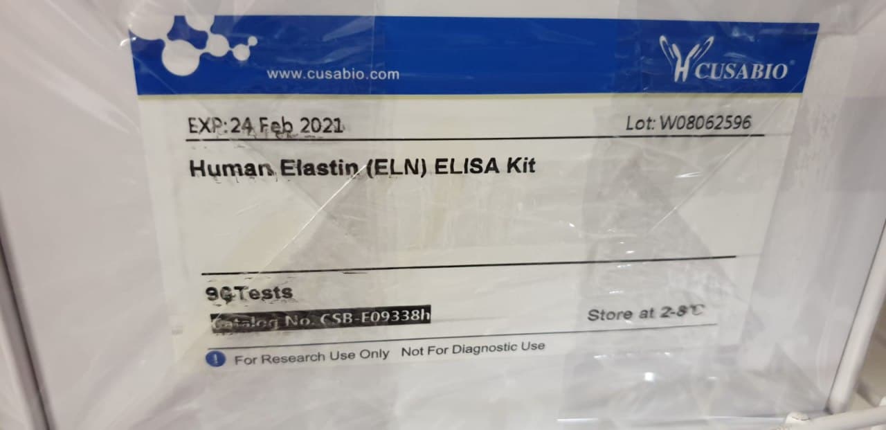
Diagnostic Accuracy of a Direct Panfungal Polymerase Chain Reaction Assay Performed on Stained Cytology Slides

Diagnostic Accuracy of a Direct Panfungal Polymerase Chain Reaction Assay Performed on Stained Cytology Slides

Molecular strategies are more and more being utilized to stained cytology slides for the analysis of neoplastic and infectious ailments. Such strategies for the identification of fungi from stained cytology slides haven’t but been evaluated. This research aimed to evaluate the diagnostic accuracy of direct (with out nucleic acid isolation) panfungal polymerase chain response (PCR) adopted by sequencing for identification of fungi and oomycetes on stained cytology slides from canines, cats, horses, and different species. Thirty-six circumstances had been recognized with cytologically identifiable fungi/oomycetes and concurrent identification through fungal tradition or immunoassay.

Twenty-nine controls had been recognized with no cytologically or histologically seen organisms and a concurrent adverse fungal tradition. Direct PCR concentrating on the inner transcribed spacer area adopted by sequencing was carried out on one cytology slide from every case and management, and the sensitivity and specificity of the assay had been calculated. The sensitivity of the panfungal PCR assay carried out on stained cytology slides was 67% total, 73% excluding circumstances with oomycetes, and 86% when contemplating solely slides with plentiful fungi. The specificity was 62%, which was attributed to amplification of fungal DNA from management slides with no seen fungus and adverse tradition outcomes.

Workplace-collected blood spots deposited on filter paper had been analysed with multiplexed affinity-based protein assays and located to be appropriate for proteomics evaluation. The protein extension assay (PEA) was used to characterize 92 proteins utilizing 1.2 mm punches in repeated samples collected from 20 employees. Overall, 97.8% of the samples and 91.3% of the analysed proteins handed high quality management. Both inside and between spot correlations utilizing six replicates from the identical particular person had been above 0.99, suggesting that comparable ranges are obtained from a number of punches from the identical spot and from consecutive spots.

Protein ranges from dried blood and moist serum from the identical people had been in contrast and the bulk of the analysed proteins had been discovered to be considerably correlated. These outcomes open up for simplified pattern assortment of blood in discipline circumstances for proteomic evaluation, but additionally spotlight that not all proteins might be robustly measured from dried complete blood. Direct panfungal PCR is succesful of offering genus- or species-level identification of fungi from stained cytology slides. Given the potential of panfungal PCR to amplify contaminant fungal DNA, this assay needs to be carried out on slides with seen fungi and interpreted along side morphologic evaluation by a medical pathologist.

Paper-based loop-mediated isothermal amplification and lateral stream (LAMP-LF) assay for identification of tissues of cattle origin

The current research was made with the aims of growth and standardization of cattle particular paper-based loop-mediated isothermal amplification cum lateral stream assay (LAMP-LFA), as a Point-of-care check (POCT) for identification of tissue of cattle origin. The parts of standardized LAMP response using cattle particular primer units had been lyophilized over paper buttons, recognized finest because the provider of LAMP reagents. Based on possible LAMP amplicon, a pair of probes was designed, tagged and its hybridization with the amplified product of paper LAMP response was optimized. The parts of lateral stream assay for detection of probe hybridized LAMP merchandise had been standardized. Analysis of profitable amplification was made by utilizing HNB dye, LAMP-LFA strip, and likewise by the standard ladder-like sample on gel electrophoresis.

The assay was discovered extremely particular for cattle with an analytical sensitivity of 0.1 pg of absolute DNA. Laboratory validation carried out on samples from completely different people of cattle, coded samples, binary meat admixture, and heat-processed cattle tissues substantiated the accuracy of the assay. Comparison with pre-standardized species-specific PCR assay taken as gold requirements revealed 100% conformity. The discipline utility of the developed assay was additional established by its compatibility with the business package eliminating the prolonged DNA extraction step and storage stability of LAMP reagent provider buttons for Four months below refrigeration. Thus, the developed assay succesful of the end result inside Three h in resource-limited settings can be utilized as POCT for identification of tissue of cattle origin.

 Diagnostic Accuracy of a Direct Panfungal Polymerase Chain Reaction Assay Performed on Stained Cytology Slides

High-Speed Lens-Free Holographic Sensing of Protein Molecules Using Quantitative Agglutination Assays

Accurate, cost-effective, easy-to-use, and point-of-care sensors for protein biomarker ranges are necessary for illness diagnostics. A cheap and compact readout method that has been used for a number of diagnostic functions is lens-free holographic microscopy, which offers an ultralarge discipline of view and submicron decision when it’s coupled with pixel super-resolution strategies. Despite its potential as a diagnostic method, lens-free microscopy has not beforehand been utilized to quantitative protein molecule sensing in answer, which might simplify sensing protocols and finally allow measurements of binding kinetics in physiological circumstances.

Here, we sense interferon-γ (an immune system biomarker) and NeutrAvidin molecules in answer by combining lens-free microscopy with a one-step bead-based agglutination assay, enabled by a customized high-speed light-emitting diode (LED) array and automatic picture processing routines. We name this a quantitative large-area binding (QLAB) sensor.

[Linking template=”default” type=”products” search=”Red Active Caspase-9 Staining Kit 25 assays” header=”1″ limit=”149″ start=”2″ showCatalogNumber=”true” showSize=”true” showSupplier=”true” showPrice=”true” showDescription=”true” showAdditionalInformation=”true” showImage=”true” showSchemaMarkup=”true” imageWidth=”” imageHeight=””]

The high-speed gentle supply offers, for the primary time, pixel super-resolved imaging of >104 2 μm beads in answer present process Brownian movement, with out vital movement blur. The automated picture processing routines allow the counting of particular person beads and clusters, offering a quantitative sensor readout that relies upon on each bead and analyte concentrations. Fits to the chemical binding idea are offered. For NeutrAvidin, we discover a restrict of detection (LOD) of <27 ng/mL (450 pM) and a dynamic vary of 2-Four orders of magnitude. For mouse interferon-γ, the LOD is <Three ng/mL (200 pM) and the dynamic vary is at the very least Four orders of magnitude. The QLAB sensor holds promise for point-of-care functions in low-resource communities and the place protocol simplicity is necessary.

Lysosome Targeting RedGreen-assay: Selective Autophagy Sensing Assay for Mammalian Cells

Lysosome Targeting RedGreen-assay: Selective Autophagy Sensing Assay for Mammalian Cells

The means of autophagy is a necessary mobile mechanism, required to take care of basic cell well being by the elimination of dysfunctional organelles, such because the ER, peroxisomes and mitochondria, in addition to protein aggregates, and micro organism. Autophagy is an especially dynamic course of, and instruments are continually being developed to check the varied steps of this course of. This protocol particulars a technique to check the top steps of autophagy-lysosomal fusion and the formation of the autolysosome. Many strategies have been used to check the varied steps of the autophagy course of.

Here we describe the RedGreen-assay (RG-assay), an immunofluorescence-based method used to visualise the concentrating on of substrates to the autolysosome in reside cells. This method takes benefit of the low lysosomal pH and over-expression of a tandem GFP-mCherry tagged protein focused to an organelle of curiosity. While within the impartial cytosol or autophagosome, each GFP and RFP will fluoresce. However, inside the autolysosome, the GFP sign is quenched as a result of low pH surroundings and the RFP emission sign will predominate. This method is quickly quantifiable and amenable to excessive throughput experiments. Additionally, by tagging the GFP-RFP tandem fluorescent protein with organelle particular concentrating on sequences, it may be used to measure a variety of substrates of autophagy.

Quantifying the ratio of alternatively spliced mRNA variants of genes with identified various splicing variants is extremely related for many purposes. Herein, we describe the validation of a quantitative PCR design for the simplified quantification of identified mRNA splice variants. The assay makes use of a single-common primer pair, twin probe design for the willpower of splicing variants in a single nicely configuration. We used murine XBP-1 splicing variants, XBP-1S and XBP-1U, to validate and show the efficiency traits of this method. Using artificial XBP-1S and XBP-1U cDNA in addition to cDNA synthesized from mouse beta-cell line MIN6, we established the efficiency parameters and dynamic vary of the assay. Reliable quantification of each variants at various focus gradients was proven.

Platelet Isolation and Activation Assays

Platelets regulate hemostasis and are the important thing determinants of pathogenic thrombosis following atherosclerotic plaque rupture. Platelets flow into in an inactive state, however develop into activated in response to wreck to the endothelium, which exposes thrombogenic materials resembling collagen to the blood circulate. Activation leads to plenty of responses, together with secretion of soluble bioactive molecules through the discharge of alpha and dense granules, activation of membrane adhesion receptors, launch of microparticles, and externalization of phosphatidylserine.

These processes facilitate agency adhesion to websites of damage and the recruitment and activation of different platelets and leukocytes, leading to aggregation and thrombus formation. Platelet activation drives the hemostatic response, and likewise contributes to pathogenic thrombus formation. Thus, quantification of platelet-associated responses is vital to many pathophysiologically related processes. Here we describe protocols for isolating, counting, and activating platelets, and for the fast quantification of cell floor proteins utilizing circulate cytometry.

Extracellular vesicles (EVs) are produced by all domains of life together with Bacteria, Archaea and Eukarya. EVs are important for mobile physiology and comprise assorted cargo: virulence components, cell wall transforming enzymes, extracellular matrix parts and even nucleic acids and metabolites. While varied protocols for isolating EVs have been established for mammalian cells, the sphere is actively growing instruments to check EVs in different organisms.

In this protocol we describe our strategies to carry out density gradient purification of EVs in bacterial cells, permitting for separation of EV subpopulations, adopted by safety assays for EV cargo characterization. Furthermore, we devised a protocol which includes a fluorescent conjugate of fatty acids into EVs, the primary to permit reside-cell EV monitoring to look at launch of EVs, together with throughout an infection of mammalian cells by pathogenic micro organism. These protocols are highly effective instruments for EV researchers as they allow the statement of EV launch and the research of the mechanisms of their formation and launch.

 Lysosome Targeting RedGreen-assay: Selective Autophagy Sensing Assay for Mammalian Cells

Ex vivo Drosophila Wing Imaginal Disc Culture and Furin Inhibitor Assay

Furin is an evolutionarily conserved proprotein convertase (PC) household enzyme with a broad vary of substrates which might be important for developmental, homeostatic, and illness pathways. Classical genetic approaches and in vitro biochemical or cell organic assays recognized that precursor types of most development issue household proteins are processed by Furin. To quantitatively assess the potential function of Furin in cleaving and modulating intercellular dispersion of a Drosophila signaling protein, we developed a easy assay by combining genetics, ex vivo organ tradition, pharmacological remedy, and imaging analyses.
[Linking template=”default” type=”products” search=”Caspase-9 Colorimetric Assay Kit – 25 assays” header=”3″ limit=”154″ start=”2″ showCatalogNumber=”true” showSize=”true” showSupplier=”true” showPrice=”true” showDescription=”true” showAdditionalInformation=”true” showImage=”true” showSchemaMarkup=”true” imageWidth=”” imageHeight=””]
The protocol herein describes easy methods to ex vivo tradition Drosophila wing imaginal discs expressing a fluorescently tagged Drosophila Fibroblast Growth Factor (FGF, Branchless/Bnl) over an extended time period within the presence of Furin inhibitors and monitor the cleavage and intercellular dispersion of the truncated Bnl components utilizing microscopy. Although the assay described right here is for assessing the impact of Furin inhibition on Bnl cleavage within the Drosophila larval wing imaginal disc, the precept and methodology can simply be adopted for every other alerts, tissue techniques, or organisms. This technique and protocol present an assay for analyzing Furin exercise on a particular substrate by straight visualizing the spatiotemporal distribution of its truncated components in an ex vivo-cultured organ.

A Highly Sensitive, Reproducible Assay for Determining 4-hydroxynonenal Protein Adducts in Biological Material

A Highly Sensitive, Reproducible Assay for Determining 4-hydroxynonenal Protein Adducts in Biological Material

Oxidative stress is related to quite a few illnesses, and markers of oxidative stress in organic materials have gotten a mainstay of each experimental and scientific/epidemiological analysis. Lipid peroxidation is a serious type of oxidative stress, however resulting from their speedy degradation and instability, lipid peroxides are notoriously tough to measure, significantly in organic specimens the place their manufacturing and removing are constantly occuring. Thus, a generally used surrogate marker of lipid peroxidation is protein adducts of 4-Hydroxynonenal (HNE), an α, β-unsaturated hydroxyalkenal (i.e., a reactive aldehyde) fashioned through degradation of oxidized polyunsaturated fatty acids (PUFAs).

HNE adducts will be measured through commercially-available immunosorbent assays, however these have their limitations resulting from extreme prices, and reproducibility amongst laboratories is difficult resulting from variability in assay sensitivity, process, and reagents. Here we current a reproducible, facile, and economically conservative protocol for quantifying HNE protein adducts. The key to this protocol is to generate HNE-adduct requirements by incubating bovine serum albumin (BSA) with HNE. These requirements are then adsorbed to immunsorbent plastic in a multi-well plate format alongside organic samples.

An enzyme-linked immunosorbent assay (ELISA) is then carried out on the multi-well plate utilizing commercially-available major and secondary antibodies, and a peroxide-based fluorescent growing reagent. This protocol is very delicate and provides benefits to business sources in that it permits for reproducible, high-throughput quantitation of HNE adducts in a lot of samples. As such, it might be helpful as a biomarker of power oxidative stress for experimental and scientific research.

Understanding the molecular mechanism governing the higher-order regulation of actin dynamics requires chemically-defined and quantitative assays. Recently, the membrane reworking giant GTPase, dynamin, has been recognized as a brand new actin cross-linking molecule. Dynamin regulates actin cytoskeleton via binding to, self-assembling round, and aligning them into actin bundles. Here we make the most of dynamin for example and current a easy protocol to investigate the actin bundling exercise in vitro. This protocol particulars the strategy for F-actin reconstitution in addition to quantitative and qualitative analyses for actin bundling exercise of dynamins. Measurement of the actin bundling exercise of different actin-binding proteins might also be utilized to this protocol with acceptable changes relying on the protein of curiosity.

Genetic identification of medicinally used Salacia species by nrDNA ITS sequences and a PCR-RFLP assay for authentication of Salacia-related well being meals

The roots and stems of a number of Salacia species have been used as conventional medicines, particularly in Ayurvedic medical system for the therapy of diabetes, rheumatism, gonorrhea, amenorrhea, pores and skin illnesses, and so forth. Due to reported proof supporting Salacia’s useful results in early-stage diabetes and different lifestyle-related illnesses, Salacia-based dietary dietary supplements and well being meals have been gaining reputation in Japan and different nations in current years. However, because of the morphological similarities between Salacia crops, significantly in the medicinally used elements (roots and stems), the authentication of the botanical identities of Salacia-derived merchandise is difficult. This research goals to develop a genetic strategy to authenticate the medicinally used Salacia species and to find out the botanical sources of the commercially obtainable Salacia-derived merchandise.
 The sequences of nuclear DNA inside transcribed spacer (ITS) and chloroplast trnK-rps16 area had been decided and in contrast between 10 plant specimens from three medicinally used Salacia species in addition to 48 samples of business crude medicine. Moreover, a PCR-restriction fragment size polymorphism (RFLP) assay was developed for speedy identification primarily based on the ITS sequences. The plant specimens from the three medicinally used Salacia species confirmed three foremost kinds of sequences in each ITS (sorts I, II, III) and trnK-rps16 (i, ii, iii) areas.
Combined the sequences of ITS and trnK-rps16 areas, S. reticulata and S. oblonga had sort I-i and kind III-iii or comparable sequences, respectively. S. chinensis had sort II-ii or II(536M)-i sequences. Forty-eight samples of business crude medicine had been recognized primarily based on ITS and trnK-rps16 DNA barcode. A handy PCR-RFLP assay utilizing Cac8I restriction enzyme was established and utilized to determine the botanical sources of well being meals merchandise bought from on-line retailers. All the twelve samples had been recognized as S. chinensis.
The nrDNA ITS sequences offered helpful info to authenticate Salacia species and to elucidate the phylogenetic relationship inside the Salacia genus. Genetic identification outcomes revealed that S. chinensis and S. reticulate are the most important sources of commercially obtainable Salacia-products. Based on the ITS sequences, a handy PCR-RFLP assay was established for the identification of the medicinally used Salacia species in addition to their derived well being meals merchandise.
 A Highly Sensitive, Reproducible Assay for Determining 4-hydroxynonenal Protein Adducts in Biological Material

The Ring-Pull Assay for Mechanical Properties of Fibrous Soft Tissues – An Analysis of the Uniaxial Approximation and a Correction for Nonlinear Thick-Walled Tissues

The ring-pull check, the place a hoop of tissue is hooked through two pins and stretched, is a well-liked biomechanical check, particularly for small arteries. Although handy and dependable, the ring check produces inhomogeneous pressure, making willpower of fabric parameters non-trivial. To decide correction elements between ring-pull-estimated and true tissue properties.  A finite-element mannequin of ring pulling was constructed for a pattern with nonlinear, anisotropic mechanical conduct typical of arteries. The pin power and pattern cross-section had been used to compute an obvious modulus at small and huge pressure, which had been in comparison with the desired properties. The ensuing corrections had been validated with experiments on porcine and ovine arteries.
[Linking template=”default” type=”products” search=”Alkaline Phosphatase Activity Fluorometric Assay Kit 500 assays” header=”2″ limit=”144″ start=”3″ showCatalogNumber=”true” showSize=”true” showSupplier=”true” showPrice=”true” showDescription=”true” showAdditionalInformation=”true” showImage=”true” showSchemaMarkup=”true” imageWidth=”” imageHeight=””]
The correction was additional utilized to experiments on mouse aortic rings to find out materials and failure properties. Calculating pressure primarily based on centerline stretch somewhat than inner-wall or outer-wall stretch afforded higher estimation of tissue properties. Additional correction elements had been developed primarily based on ring wall thickness H, centerline ring radius R c , and pin radius a. The corrected estimates for tissue properties had been in good settlement with uniaxial stretch experiments.

Gentamicin Protection Assay to Determine the Number of Intracellular Bacteria during Infection of Human TC7 Intestinal Epithelial Cells by Shigella flexneri

Gentamicin Protection Assay to Determine the Number of Intracellular Bacteria during Infection of Human TC7 Intestinal Epithelial Cells by Shigella flexneri

Shigella flexneri is an intracellular bacterial pathogen that positive aspects entry to the intestine epithelium utilizing a specialised Type III Secretion System (T3SS). Various determinants mediating this invasive an infection have been experimentally verified utilizing the classical gentamicin safety assay introduced right here. In this assay epithelial cell strains are contaminated by micro organism in vitro and the extracellular micro organism are killed by gentamicin. The internalized micro organism, that are shielded from the bactericidal motion of gentamicin, are recovered by lysing the epithelial cells and enumerated by figuring out the colonies fashioned on strong medium.

Various methods primarily based on gentle microscopy, reminiscent of immunofluorescence and micro organism expressing fluorescent proteins, are additionally used for finding out intracellular micro organism. However, these methods should not solely labor intensive and require refined tools, however principally are additionally not quantitative. Despite being a straightforward quantitative technique to research invasiveness of micro organism, the gentamicin safety assay can’t distinguish between the survival and multiplication of the internalized micro organism over longer incubation durations.

To alleviate the problems created by multiplication and dissemination of internalized micro organism, complementary assays like plaque formation assays are required. This protocol presents a straightforward and cost-effective technique to decide the invasiveness and the capability to set up an an infection of Shigella below totally different circumstances. Automatic and visible interpretation of outcome bands have been additionally in contrast for the immunochromatography-based BinaxNOW and ImmuView UATs.

Urinary antigen exams (UATs) are sometimes used to diagnose Legionnaires’ illness as they’re speedy and simple to carry out on readily obtainable urine samples with out the want for specialised abilities in contrast to standard strategies. Recently developed automated readers for UATs could present goal outcomes interpretation, particularly in instances of weak outcome bands. Using 53 outlined affected person urine samples, we evaluated the efficiency of the BinaxNOW Legionella Antigen Card (Abbott), ImmuView S. pneumoniae and Legionella (SSI Diagnostica), STANDARD F Legionella Ag FIA (SD Biosensor), and Sofia Legionella FIA (Quidel) concurrently with their respective automated readers.

SARS-CoV-2 lateral circulation assays for attainable use in nationwide covid-19 seroprevalence surveys (React 2): diagnostic accuracy research

ensitivity analyses have been carried out on sera saved from 320 earlier contributors in the React 2 programme with confirmed earlier extreme acute respiratory syndrome coronavirus 2 (SARS-CoV-2) an infection. Specificity analyses have been carried out on 1000 prepandemic serum samples. 100 new contributors with confirmed earlier SARS-CoV-2 an infection attended research clinics for finger prick testing.  Laboratory sensitivity and specificity analyses have been carried out for seven LFIAs on a minimal of 200 serum samples from contributors with confirmed SARS-CoV-2 an infection and 500 prepandemic serum samples, respectively.
Three LFIAs have been discovered to have a laboratory sensitivity superior to the finger prick sensitivity of the LFIA presently utilized in React 2 seroprevalence research (84%). These LFIAs have been then additional evaluated by way of finger prick testing on contributors with confirmed earlier SARS-CoV-2 an infection: two LFIAs (Surescreen, Panbio) have been evaluated in clinics in June-July 2020 and the third LFIA (AbC-19) in September 2020. A spike protein enzyme linked immunoassay and hybrid double antigen binding assay have been used as laboratory reference requirements.
The accuracy of LFIAs in detecting immunoglobulin G (IgG) antibodies to SARS-CoV-2 in contrast with two reference requirements. The sensitivity and specificity of seven new LFIAs that have been analysed utilizing sera diverse from 69% to 100%, and from 98.6% to 100%, respectively (in contrast with the two reference requirements). Sensitivity on finger prick testing was 77% (95% confidence interval 61.4% to 88.2%) for Panbio, 86% (72.7% to 94.8%) for Surescreen, and 69% (53.8% to 81.3%) for AbC-19 in contrast with the reference requirements. Sensitivity for sera from matched medical samples carried out on AbC-19 was considerably larger with serum than finger prick at 92% (80.0% to 97.7%, P=0.01). Antibody titres diverse significantly amongst cohorts. The numbers of constructive samples recognized by finger prick in the lowest antibody titre quarter diverse amongst LFIAs.
Gentamicin Protection Assay to Determine the Number of Intracellular Bacteria during Infection of Human TC7 Intestinal Epithelial Cells by Shigella flexneri

Establishment of Novel Protein Interaction Assays between Sin3 and REST Using Surface Plasmon Resonance and Time-Resolved Fluorescence Energy Transfer

Repressor element-1 (RE-1) or neural restrictive silencer component (NRSE) certain with a zinc finger transcription repressor, RE-1 silencing transcription issue (REST, also called neural restrictive silencer issue, NRSF) has been recognized as a basic repressor component in lots of genes, together with neuronal genes. Genes regulated by REST/NRSF regulate multifaceted neuronal phenotypes, and their defects in the equipment trigger neuropathies, issues of neuron exercise), autism and so forth. In REST repressions, the N-terminal repressor area recruits Sin3B by way of its paired amphipathic helix 1 (PAH1) area, which performs an necessary function as a scaffold for histone deacetylase 1 and a pair of. This equipment has a important function in sustaining neuronal robustness.

[Linking template=”default” type=”products” search=”NADH Fluorometric Assay Kit 100 assays” header=”2″ limit=”132″ start=”4″ showCatalogNumber=”true” showSize=”true” showSupplier=”true” showPrice=”true” showDescription=”true” showAdditionalInformation=”true” showImage=”true” showSchemaMarkup=”true” imageWidth=”” imageHeight=””]

In this research, so as to set up protein-protein interplay assays mimicking a binding floor between Sin3B and REST, we chosen necessary amino acids from structural data of the PAH1/REST complicated after which tried to reconstitute it utilizing recombinant quick peptides derived from PAH1/REST. Initially, we validated whether or not biotinylated REST interacts with glutathione S-transferase (GST)-tagged PAH1 and whether or not one other PAH1 peptide (PAH1-FLAG) competitively binds with biotinylated REST utilizing floor plasmon resonance (SPR). We noticed a direct interplay and aggressive binding of two PAH1 peptides.

Secondly, so as to set up a high-throughput and high-dynamic-range assay, we utilized an simply carried out novel time-resolved fluorescence vitality switch (TR-FRET) assay, and intently monitored this interplay. Finally, we succeeded in establishing a novel high-quality TR-FRET assay and a novel interplay assay primarily based on SPR.

SARS-CoV-2 diagnosis: What is new?

How do tests that reveal the SARS-CoV-2 virus work?

Covid-19 diagnostic tests are an essential tool for monitoring the spread of the pandemic. The World Health Organization (WHO) has repeatedly asked health agencies to make coronavirus testing a top priority, but international response has been mixed.

Why does their use vary so much from one country to another?

Thanks to a combination of enabling regulations and logistical readiness, some countries, such as South Korea and Singapore, have been able to rapidly deploy massive test campaigns targeting thousands of individuals; but elsewhere, several countries have struggled to keep pace.

What are the new types of tests in preparation?

Meanwhile, in the United States, faced with delays of several weeks from hospitals and manufacturers, university research laboratories have developed their diagnostic tools to meet needs.

Test, instructions for use

Most Covid-19 tests are now performed on viral genetic material from samples from the nose or pharynx. They are based on a molecular biology tool designated by the acronym RT-PCR. PCR (for polymerase chain reaction) is a method of duplicating (or amplifying) a DNA sequence in very large numbers thanks to an enzyme, the polymerase. We can then analyze the DNA.

However, since the coronavirus genome is in the form of RNA, it must first be transformed into DNA, which another enzyme does, reverse transcriptase (RT).

Amplification starts from short sequences, added primers, complementary to the sequence to be duplicated. However, PCR can only detect the virus when it is present, and is of no use when the epidemic is resolved. What is more, it sometimes produces false positives when the reagents (in particular the primers and the products necessary for the duplication of DNA) are contaminated.

Laboratories around the world quickly adapted their RT-PCR tests to SARS-CoV-2, by designing appropriate primers, complementary to various fragments of the genetic sequence of the virus.

Which countries have been reactive?

Several countries have been able to test very large numbers of people (see figure below). South Korea reacted when the first cases appeared at the end of January by deploying large quantities of rapid tests and then monitoring those in contact with those who had tested positive. Regulations promulgated after the Middle East respiratory syndrome (MERS) epidemic in 2015 (with 186 cases and 35 deaths, South Korea was the second most affected country after Saudi Arabia which counted 688 cases and 282 deaths) allowed rapid release of a version of the WHO test, and four companies produced enough to test up to 10,000 people per day.

In this way, explains Angela Rasmussen of Columbia University in New York, the South Korean authorities were able to identify the slightly ill and asymptomatic people and prevent them from spreading the virus. “The country has not had to take the drastic social distancing measures as in other countries,” she added.

As for Singapore, the city-state developed a test as soon as the coronavirus appeared in China, explains Vernon Lee, of the Singapore Ministry of Health, and was very early in battle order to track down the cases. Hospital laboratories were quickly given the green light to use other authorized PCR tests, provided that the results of the first cohort were validated by a national laboratory. They also focused the tests on the most essential cases such as patients with pneumonia or people at high risk, for example elderly people.

College of public health and occupational physicians
-Singapore-

Essential logistics were installed after the appearance in 2003 of another coronavirus, the SARS-Cov: it notably provides sufficient capacities in laboratories, trained personnel, reagents…

“In Singapore, we are ready and have ” a medical and hospital system strengthened since the SARS episode. It’s not something we created because of the Covid-19, ”

Vernon Lee.

The latecomers

Health experts have criticized the United States for its slow response to the pandemic, in particular the low availability of tests and their slow implementation. According to specialists, these failures allowed the virus to spread without being detected and imposed strict social distancing measures to contain it.

US health officials have admitted their fault. At a congressional hearing on March 12, the director of the National Institute of Allergies and Infectious Diseases (NIAID), Anthony Fauci, said that the country “is unable” to test the population and was not in able to meet demand for testing.

Kenneth Bernard, a former adviser to several US presidents on biodefense, said a coordinator or a White House office responsible for responding to the pandemic could have facilitated communication between the agencies and drawn up a plan to intensify the tests. But the person who held the office left President Donald Trump’s National Security Council … in May 2018.

“We had a problem with the scale. It is one thing to do a benchmark test at the CDC (Center for Disease Control and Prevention), but another to do millions of them, “warns Kenneth Bernard.

Rigid regulations for new diagnostic tests have been an additional barrier. Initially, the only approved American test was created by the CDC, but it was found to be defective. And only public health services were allowed to use it. The Food and Drug Administration (FDA) did not relax these rules until the end of February and did not allow private labs to test until mid-March. It was only on the 21st that the US authorities authorized a rapid test that did not require samples to be sent to a laboratory. It should be ready soon.

With the federal response long overdue, the virology laboratories at the University of Washington in Seattle organized the first major national testing operation. The researchers adapted the WHO protocol to work with the reagents and platforms they had on hand. Dozens of other laboratories and research consortia have followed suit.

“The federal government made the decision to severely limit access [to the tests] at first,” said Joshua Sharfstein, a former FDA official. “If you could go back and tell the FDA in mid-January that you’re really going to need a million tests soon enough, they would have chosen a different strategy. “

What about serological tests?

One of the main goals is to develop a serological test, which is one that can detect past viral infections by looking for antibodies that a formerly infected person has produced to fight the virus. Such a test would reveal the extent of viral spread in a population and would provide useful public health information.

“For the moment, it is clear that we are only seeing the tip of the iceberg: patients so severely affected that they must be hospitalized or even receive intensive care there,” explains Robert Garry, virologist at Tulane University, in New Orleans, Louisiana. “What is worrying is that there are a lot of patients with little or even asymptomatic disease outside. “

Several groups, including that of Robert Garry, are working on carrying out such a serological test. Another team, including researchers from Icahn School of Medicine at Mount Sinai Hospital in New York, developed one that she describes in a prepublication, not peer reviewed, posted on medRxiv on March 18. “We don’t see any obstacle to carrying out these tests, only a few adjustments,” says Robert Garry.

A team in Singapore used serological tests to trace contacts of patients, but at the time, the method used had not been validated for clinical use. “We think this is the first time in the world that this type of testing has been used in this context,” said Danielle Anderson, virologist at the faculty of medicine at Duke and Singapore Universities, in Singapore, during a press conference in February.

Make way for innovation

Research groups led by two of CRISPR’s leading specialists, the tool that makes it easy to modify an organism’s genome, are each working on tests that leverage this gene editing technique to improve testing.

First, at the University of Washington, virologist Keith Jerome and his colleagues improved the SHERLOCK test (for specific high sensitivity enzymatic reporter unlocking) developed by Feng Zhang at the Massachusetts Institute of Technology. The latter said he had sent some 1,600 test kits to ten laboratories around the world.

Next, Mammoth Biosciences, a California-based biotechnology company co-founded by Jennifer Doudna of the University of California at Berkeley, is working on another CRISPR-based method called DETECTR to detect SARS-CoV-2.

Both techniques use CRISPR’s ability to recognize and cut specific genetic sequences. Here, he also cuts a reporter molecule added to the reaction, which relatively quickly reveals the presence of viral genetic material.

“Whenever we are faced with an epidemic, we are late because we do not have rapid diagnostic tools to detect and identify the new organism in question,” said Charles Chiu of the University of California at San Francisco, who works with Mammoth Biosciences. “The main advantages of CRISPR are its specificity and speed, a test which can be carried out in 5 to 10 minutes. “

Robert Garry, at Tulane, is optimistic about such advances. “It is a powerful technique. I’ve seen it work with the Zika virus and a few other viruses, “he says. “If anyone can find a way to lessen the impact of the Covid-19, let’s do it. “