NCI-H226 [H226] Cell Line: A Deep Dive into Its Role in Lung Cancer Research

Introduction

Lung cancer remains a leading cause of cancer-related deaths worldwide, with mesothelioma and non-small cell lung cancer (NSCLC) being particularly aggressive subtypes (National Cancer Institute). Among the available in vitro models, the NCI-H226 [H226] cell line is widely used to study lung cancer biology, molecular pathways, and therapeutic responses.

Origin and Establishment of NCI-H226 Cells

The NCI-H226 cell line was derived from a pleural effusion of a patient with squamous cell carcinoma, a subtype of NSCLC (Cellosaurus Database). Developed at the National Cancer Institute (NCI), it has since been extensively utilized in research studies to understand tumor progression, metastasis, and resistance mechanisms (PubMed).
Genetic and Molecular Characteristics

NCI-H226 cells exhibit genetic alterations commonly found in lung cancer, including:

  • TP53 Mutation: A crucial mutation affecting tumor suppression, leading to unchecked cell proliferation (NCBI).
  • EGFR Wild-Type Status: Unlike some NSCLC lines, H226 lacks EGFR mutations, making it a model for studying non-EGFR-driven lung cancer (Cancer Genome Atlas).
  • MET Amplification: This genetic alteration contributes to tumor growth and resistance to certain therapies (NIH).
  • PTEN Alterations: PTEN loss has been associated with increased aggressiveness in lung cancer (PubMed).

Cellular and Morphological Features

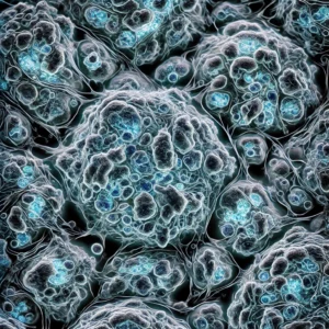
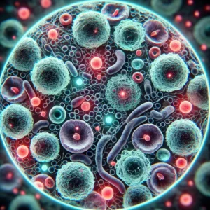
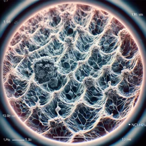
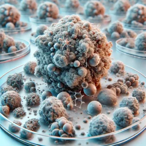
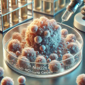
Microscopic analysis reveals that NCI-H226 cells are adherent with an epithelial-like morphology. They form monolayers in culture, closely mimicking squamous cell carcinoma tumors (ATCC).

Culture and Growth Conditions

For optimal growth, NCI-H226 cells require the following conditions:

  • Medium: RPMI-1640 supplemented with 10% fetal bovine serum (FBS – Heat Inactivated Qualified Imported Fetal Bovine Serum, USDA Approved)
  • Atmosphere: 37°C with 5% CO2
  • Doubling Time: Approximately 36-48 hours (DSMZ)
  • Adherent Nature: Cells require proper attachment substrates to grow efficiently.
  • Passaging Recommendations: Subculturing is recommended at 80-90% confluency to maintain optimal growth conditions.
  • Cryopreservation: Cells should be frozen in 90% FBS and 10% DMSO for long-term storage.
Applications in Lung Cancer Research

The NCI-H226 cell line plays a critical role in cancer studies, with broad applications including:

1. Tumor Progression and Metastasis

Researchers use this model to explore molecular pathways driving squamous cell carcinoma progression, particularly transcriptional factors regulating tumor invasion (PubMed).

2. Drug Screening and Resistance Mechanisms

NCI-H226 is used to evaluate chemotherapy and targeted therapy responses, particularly in cisplatin-based treatments and resistance studies (NIH).

3. Radiation Sensitivity Studies

This cell line has been employed in studies examining radiation therapy effects on squamous cell lung cancer, aiding in personalized treatment strategies (Cancer.gov).

4. Immunotherapy Research

As immunotherapy becomes a standard treatment for lung cancer, NCI-H226 helps assess the efficacy of checkpoint inhibitors like PD-1/PD-L1 blockers (NCI).

5. Gene Expression and Epigenetic Studies

Scientists use NCI-H226 to analyze epigenetic modifications such as DNA methylation and histone modifications, which play significant roles in lung cancer development (NIH Epigenetics Program).

6. Co-Culture Systems and Microenvironment Studies

Recent advancements have enabled the use of NCI-H226 in co-culture systems with fibroblasts, immune cells, and endothelial cells to mimic the tumor microenvironment (NCBI).

7. Organoid and 3D Culture Models

Researchers are developing organoid models derived from NCI-H226 to study drug responses in a more physiologically relevant setting (NIH 3D Models Program).

Challenges and Limitations

Despite its importance, the NCI-H226 cell line has certain limitations:

  • Limited Representation of Tumor Heterogeneity: Single-cell models may not fully capture patient tumor diversity (Cancer.gov).
  • Lack of Immune Components: Like most in vitro models, H226 does not include immune interactions, which are crucial in modern therapies (NIH Immuno-Oncology Studies).
  • Variable Drug Sensitivity: Different subclones of H226 may exhibit varied responses to drugs, making reproducibility a challenge (PubMed).
  • Genetic Drift Over Passages: Extended culturing may result in genetic and phenotypic drift, affecting experimental reproducibility (NCBI).
Future Directions

To enhance its translational value, researchers are integrating 3D organoid cultures, co-culture systems with stromal cells, and CRISPR gene-editing techniques to better replicate patient tumors (NCBI). Additionally, single-cell RNA sequencing is being applied to explore intra-tumoral heterogeneity in greater detail (NIH).

Conclusion

NCI-H226 is a crucial tool for lung cancer research, offering valuable insights into tumor biology, drug resistance, radiation sensitivity, and therapeutic efficacy. With advances in genomics, cell engineering, and immunotherapy, this model continues to drive precision medicine and novel treatment strategies. Researchers worldwide are leveraging its unique characteristics to improve our understanding of squamous cell carcinoma and develop more effective treatment options.

For additional resources, explore the National Cancer Institute, PubMed, and NIH Clinical Trials.
 

NCI-H226 [H226] Cell Line: A Deep Dive into Its Role in Lung Cancer Research

Introduction

Lung cancer remains a leading cause of cancer-related deaths worldwide, with mesothelioma and non-small cell lung cancer (NSCLC) being particularly aggressive subtypes (National Cancer Institute). Among the available in vitro models, the NCI-H226 [H226] cell line is widely used to study lung cancer biology, molecular pathways, and therapeutic responses.

Origin and Establishment of NCI-H226 Cells

The NCI-H226 cell line was derived from a pleural effusion of a patient with squamous cell carcinoma, a subtype of NSCLC (Cellosaurus Database). Developed at the National Cancer Institute (NCI), it has since been extensively utilized in research studies to understand tumor progression, metastasis, and resistance mechanisms (PubMed).

Genetic and Molecular Characteristics

NCI-H226 cells exhibit genetic alterations commonly found in lung cancer, including:

  • TP53 Mutation: A crucial mutation affecting tumor suppression, leading to unchecked cell proliferation (NCBI).
  • EGFR Wild-Type Status: Unlike some NSCLC lines, H226 lacks EGFR mutations, making it a model for studying non-EGFR-driven lung cancer (Cancer Genome Atlas).
  • MET Amplification: This genetic alteration contributes to tumor growth and resistance to certain therapies (NIH).
  • PTEN Alterations: PTEN loss has been associated with increased aggressiveness in lung cancer (PubMed).

Cellular and Morphological Features

Microscopic analysis reveals that NCI-H226 cells are adherent with an epithelial-like morphology. They form monolayers in culture, closely mimicking squamous cell carcinoma tumors (ATCC).

Culture and Growth Conditions

For optimal growth, NCI-H226 cells require the following conditions:

  • Medium: RPMI-1640 supplemented with 10% fetal bovine serum (FBS)
  • Atmosphere: 37°C with 5% CO2
  • Doubling Time: Approximately 36-48 hours (DSMZ)
  • Adherent Nature: Cells require proper attachment substrates to grow efficiently.

Applications in Lung Cancer Research

The NCI-H226 cell line plays a critical role in cancer studies, with broad applications including:

1. Tumor Progression and Metastasis

Researchers use this model to explore molecular pathways driving squamous cell carcinoma progression, particularly transcriptional factors regulating tumor invasion (PubMed).

2. Drug Screening and Resistance Mechanisms

NCI-H226 is used to evaluate chemotherapy and targeted therapy responses, particularly in cisplatin-based treatments and resistance studies (NIH).

3. Radiation Sensitivity Studies

This cell line has been employed in studies examining radiation therapy effects on squamous cell lung cancer, aiding in personalized treatment strategies (Cancer.gov).

4. Immunotherapy Research

As immunotherapy becomes a standard treatment for lung cancer, NCI-H226 helps assess the efficacy of checkpoint inhibitors like PD-1/PD-L1 blockers (NCI).

5. Gene Expression and Epigenetic Studies

Scientists use NCI-H226 to analyze epigenetic modifications such as DNA methylation and histone modifications, which play significant roles in lung cancer development (NIH Epigenetics Program).

Challenges and Limitations

Despite its importance, the NCI-H226 cell line has certain limitations:

  • Limited Representation of Tumor Heterogeneity: Single-cell models may not fully capture patient tumor diversity (Cancer.gov).
  • Lack of Immune Components: Like most in vitro models, H226 does not include immune interactions, which are crucial in modern therapies (NIH Immuno-Oncology Studies).
  • Variable Drug Sensitivity: Different subclones of H226 may exhibit varied responses to drugs, making reproducibility a challenge (PubMed).

Future Directions

To enhance its translational value, researchers are integrating 3D organoid cultures, co-culture systems with stromal cells, and CRISPR gene-editing techniques to better replicate patient tumors (NCBI). Additionally, single-cell RNA sequencing is being applied to explore intra-tumoral heterogeneity in greater detail (NIH).

Conclusion

NCI-H226 is a crucial tool for lung cancer research, offering valuable insights into tumor biology, drug resistance, radiation sensitivity, and therapeutic efficacy. With advances in genomics, cell engineering, and immunotherapy, this model continues to drive precision medicine and novel treatment strategies. Researchers worldwide are leveraging its unique characteristics to improve our understanding of squamous cell carcinoma and develop more effective treatment options.

For additional resources, explore the National Cancer Institute, PubMed, and NIH Clinical Trials.
 

Exploring Human Pancreatic Cancer Cells: A Comprehensive Study on PaTu 8988t

Introduction

Pancreatic cancer remains one of the most aggressive and lethal malignancies, with a five-year survival rate of less than 10% (National Cancer Institute). Among the various in vitro models used to study this disease, the PaTu 8988t cell line has emerged as a valuable tool for understanding the molecular mechanisms of pancreatic adenocarcinoma and for testing potential therapeutic agents.

Origin and Establishment of PaTu 8988t Cells

PaTu 8988t cells were derived in 1985 from a liver metastasis of a primary pancreatic adenocarcinoma in a 64-year-old female patient (Cellosaurus Database). This cell line was developed alongside its sister cell line, PaTu 8988s, which originates from the same tumor but exhibits different biological characteristics. While PaTu 8988s represents a less differentiated phenotype, PaTu 8988t retains more epithelial-like features, making it an essential model for studying pancreatic cancer differentiation and metastasis (PubMed).

Genetic and Molecular Characteristics

PaTu 8988t cells display several key genetic mutations that are hallmarks of pancreatic cancer. These include:

  • KRAS Mutation: The KRAS oncogene is frequently mutated in pancreatic cancers. PaTu 8988t cells harbor a homozygous p.Gly12Val mutation, which plays a pivotal role in uncontrolled cell growth (NCBI).
  • TP53 Mutation: A homozygous p.Arg282Trp mutation in the tumor suppressor gene TP53 is present, affecting cell cycle regulation and apoptosis (Cancer Genome Atlas).
  • SMAD4 Deletion: The loss of SMAD4, a gene involved in the TGF-β signaling pathway, contributes to increased tumor aggressiveness and poor prognosis (PubMed).

Cellular and Morphological Features

Microscopic analysis of PaTu 8988t cells reveals an adherent, epithelial-like morphology with features of differentiation. When injected into nude mice, these cells form tubular structures that resemble those found in primary pancreatic adenocarcinoma, providing researchers with an excellent in vivo model for studying tumor progression (ATCC).

Culture and Growth Conditions

For optimal growth, PaTu 8988t cells require the following culture conditions:

  • Medium: Dulbecco’s Modified Eagle Medium (DMEM)
  • Serum: 5% fetal bovine serum (FBS) + 5% horse serum
  • Supplements: 2 mM L-glutamine
  • Doubling Time: 22-30 hours (DSMZ)

These cells thrive in standard 37°C incubators with 5% CO2, making them relatively easy to maintain for laboratory research.

Applications in Cancer Research

The PaTu 8988t cell line has been widely used in pancreatic cancer studies, with applications including:

1. Tumor Biology and Metastasis Research

By comparing PaTu 8988t with its sister cell line PaTu 8988s, scientists can examine differences in metastatic potential and differentiation status (PubMed).

2. Drug Screening and Therapeutic Development

Due to its well-characterized KRAS and TP53 mutations, PaTu 8988t serves as an excellent model for testing targeted therapies such as MEK inhibitors and TP53 reactivators (NIH).

3. Genetic and Epigenetic Studies

PaTu 8988t cells allow researchers to investigate the impact of epigenetic modifications and gene expression alterations in pancreatic cancer progression (National Library of Medicine).

Challenges and Limitations

While PaTu 8988t has proven to be a valuable research tool, it is essential to acknowledge its limitations:

  • Genomic Instability: Like many cancer cell lines, PaTu 8988t undergoes genetic drift over multiple passages, which may alter experimental outcomes (Cell Line Authentication Consortium).
  • Lack of Immune System Interactions: In vitro models do not fully replicate tumor-immune system interactions, which are crucial for understanding therapeutic responses (NIH Immuno-Oncology Studies).
  • Differences from Primary Tumors: Despite its high fidelity to pancreatic adenocarcinoma, cell line models may not fully capture the heterogeneity observed in human tumors (Cancer.gov).

Future Directions

To enhance the translational impact of research using PaTu 8988t, scientists are exploring 3D culture systems, patient-derived xenografts (PDX), and organoids to better mimic pancreatic tumor microenvironments (NCBI). CRISPR-Cas9 technology is also being leveraged to create genetically modified versions of PaTu 8988t, allowing for the study of specific gene functions (NIH).

Conclusion

PaTu 8988t is an essential pancreatic cancer cell line, providing researchers with an invaluable in vitro model for studying tumor biology, drug responses, and molecular signaling pathways. With ongoing advances in genomic technologies and tumor modeling, this cell line continues to drive discoveries aimed at improving pancreatic cancer diagnosis and treatment.

For more information, explore resources from National Cancer Institute and PubMed.


Recombinant Mouse Sonic Hedgehog Protein (Shh), Partial (Active): Functions, Applications, and Research Insights

Introduction to Sonic Hedgehog Protein (Shh)

The Sonic Hedgehog (Shh) protein is a critical signaling molecule involved in embryonic development, cell differentiation, and tissue regeneration. The recombinant Mouse Sonic Hedgehog protein (Shh), Partial (Active) is widely used in biomedical research to study its role in developmental biology, neurogenesis, and cancer progression.

For more information on Sonic Hedgehog signaling, visit the National Institutes of Health (NIH) (nih.gov) and the National Center for Biotechnology Information (NCBI) (ncbi.nlm.nih.gov).

Structure and Mechanism of Action

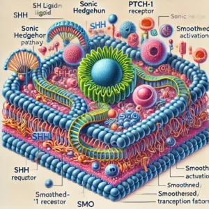
The Sonic Hedgehog (Shh) protein belongs to the Hedgehog (Hh) signaling family, which also includes Desert Hedgehog (Dhh) and Indian Hedgehog (Ihh). The active recombinant form of the protein retains its N-terminal signaling domain, which is crucial for binding to its primary receptor, Patched-1 (PTCH1). Upon binding, PTCH1 releases its inhibition on Smoothened (SMO), activating downstream transcription factors.

For further details on Hedgehog signaling pathways, explore The Cancer Genome Atlas (TCGA) (cancergenome.nih.gov) and Genomic Data Commons (GDC) (gdc.cancer.gov).

Biological Functions of Shh

1. Embryonic Development and Morphogenesis

Shh plays a fundamental role in limb patterning, neural tube formation, and organogenesis. It is particularly crucial for the development of the spinal cord, brain, and skeletal system.

To learn more about Shh’s role in development, visit National Institute of Child Health and Human Development (NICHD) (nichd.nih.gov) and Harvard Stem Cell Institute (hsci.harvard.edu).

2. Neurogenesis and Brain Function

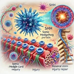
Shh signaling is involved in maintaining neural stem cells, guiding axon growth, and promoting neuroprotection. It has been studied for its role in neurodegenerative disorders such as Parkinson’s disease and Alzheimer’s disease.

For neurogenesis research, refer to National Institute of Neurological Disorders and Stroke (NINDS) (ninds.nih.gov) and Johns Hopkins Brain Science Institute (hopkinsmedicine.org).

3. Cancer Progression and Tumorigenesis

Aberrant Hedgehog signaling activation has been linked to multiple cancers, including medulloblastoma, basal cell carcinoma, and pancreatic cancer. Inhibitors targeting Shh pathway components (such as SMO inhibitors like Vismodegib and Sonidegib) have been developed as therapeutic agents.

For more details on Shh-related cancers, check National Cancer Institute (NCI) (cancer.gov) and MD Anderson Cancer Center (mdanderson.org).

Applications of Recombinant Mouse Shh Protein

1. Stem Cell Research and Regenerative Medicine

Recombinant Shh protein is commonly used to induce stem cell differentiation into neuronal, cardiac, and skeletal lineages. It plays a pivotal role in promoting tissue regeneration and repair.

For resources on stem cell applications, visit Stanford University Stem Cell Institute (med.stanford.edu) and California Institute for Regenerative Medicine (CIRM) (cirm.ca.gov).

2. Drug Development and Therapeutic Targeting

Shh inhibitors and activators are widely explored for treating cancer, neurodegenerative diseases, and tissue injuries. The development of Hedgehog pathway inhibitors has opened new avenues in oncology and regenerative medicine.

For clinical trials involving Hedgehog pathway therapies, refer to ClinicalTrials.gov (clinicaltrials.gov) and U.S. Food and Drug Administration (FDA) (fda.gov).

3. Neural Injury and Spinal Cord Repair

Studies indicate that Shh signaling enhances axonal regrowth and remyelination after spinal cord injury. This makes Shh a potential target for neuroprotective therapies.

For updates on spinal cord injury research, visit Christopher & Dana Reeve Foundation (christopherreeve.org) and National Institute of Neurological Disorders and Stroke (NINDS) (ninds.nih.gov).

Challenges and Future Directions

While recombinant Shh protein offers promising applications, several challenges remain:

  • Dosage and toxicity concerns in clinical applications.
  • Cross-talk with other signaling pathways that complicates therapeutic targeting.
  • Delivery mechanisms for effective in vivo applications.

For ongoing research, explore Massachusetts Institute of Technology (MIT) Biological Engineering (be.mit.edu) and National Human Genome Research Institute (NHGRI) (genome.gov).

Conclusion

Recombinant Mouse Sonic Hedgehog protein (Shh), Partial (Active) remains an essential tool for studying developmental biology, cancer, and regenerative medicine. Ongoing research and therapeutic advancements will further refine its applications.

For further reading, explore Mayo Clinic Research (mayoclinic.org) and Salk Institute for Biological Studies (salk.edu).

As new discoveries emerge, the Shh pathway will continue to be a key target in medical and scientific research, shaping the future of therapeutic interventions.


MDA-MB-157 Cells: A Key Model for Triple-Negative Breast Cancer Research and Therapeutic Innovations

Introduction to MDA-MB-157 Cells

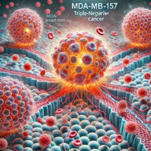
MDA-MB-157 is a human breast cancer cell line that originates from a triple-negative breast cancer (TNBC) patient. These cells are widely used in cancer research, particularly in studies focused on tumor progression, metastasis, drug resistance, and therapeutic targets. Unlike hormone receptor-positive breast cancer cells, MDA-MB-157 lacks estrogen receptor (ER), progesterone receptor (PR), and HER2 expression, making it a challenging yet significant model for studying aggressive breast cancers.

For more details on triple-negative breast cancer (TNBC), visit the National Cancer Institute (NCI) (cancer.gov) and Centers for Disease Control and Prevention (CDC) (cdc.gov).

Origin and Characteristics

MDA-MB-157 cells were originally isolated from a 43-year-old female patient with breast adenocarcinoma. These cells exhibit high metastatic potential, mesenchymal-like morphology, and significant resistance to conventional therapies. Their aggressive behavior is relevant to studies on epithelial-mesenchymal transition (EMT), invasion, and tumor microenvironment interactions.

For more information on the molecular characterization of MDA-MB-157, refer to the National Center for Biotechnology Information (NCBI) (ncbi.nlm.nih.gov).

Applications in Cancer Research

1. Tumor Progression and Metastasis

MDA-MB-157 cells provide a valuable in vitro model for studying breast cancer invasion and metastasis. Their mesenchymal-like phenotype makes them useful for analyzing migration, extracellular matrix degradation, and cancer stem cell (CSC) properties.

Research papers on breast cancer metastasis can be accessed via PubMed (pubmed.ncbi.nlm.nih.gov) and National Institutes of Health (NIH) (nih.gov).

2. Drug Resistance Studies

Triple-negative breast cancer is notorious for its lack of targeted therapies and high resistance to chemotherapy. Studies using MDA-MB-157 focus on multidrug resistance (MDR) mechanisms, apoptosis evasion, and novel therapeutic agents.

For research on drug resistance in TNBC, visit ClinicalTrials.gov (clinicaltrials.gov) and the U.S. Food and Drug Administration (FDA) (fda.gov).

3. Immunotherapy and Targeted Therapies

Since MDA-MB-157 lacks hormone receptors, researchers explore immune checkpoint inhibitors, monoclonal antibodies, and small molecule inhibitors as potential treatment strategies.

For updates on cancer immunotherapy trials, refer to National Cancer Institute (NCI) (cancer.gov) and Cancer Research UK (cancerresearchuk.org).

4. Gene Expression and Molecular Profiling

MDA-MB-157 cells are commonly analyzed for gene expression profiling and transcriptomic studies to identify oncogenic pathways and novel biomarkers.

For access to publicly available gene expression data, visit Genomic Data Commons (GDC) (gdc.cancer.gov) and The Cancer Genome Atlas (TCGA) (cancergenome.nih.gov).

5. 3D Culture Models and Organoids

Recent advances in three-dimensional (3D) culture techniques and tumor organoid models have significantly improved cancer research. MDA-MB-157 cells have been used to develop organoids that better simulate tumor biology, drug response, and microenvironment interactions.

To learn more about 3D culture technologies, visit Harvard Wyss Institute for Biologically Inspired Engineering (wyss.harvard.edu) and National Cancer Institute’s Organoid Research (cancer.gov).

Challenges and Limitations

Despite its relevance in breast cancer research, the MDA-MB-157 cell line has certain limitations:

  • High heterogeneity: Exhibits genetic instability, making reproducibility difficult.
  • Low tumorigenicity in vivo: Unlike some other TNBC cell lines, MDA-MB-157 has relatively low tumor formation capacity in xenograft models.
  • Limited therapeutic response: Poor sensitivity to standard chemotherapeutics limits its application in drug screening assays.

For a detailed review on breast cancer cell line models, explore research published by American Association for Cancer Research (AACR) (aacr.org) and National Comprehensive Cancer Network (NCCN) (nccn.org).

Future Directions

Ongoing research is aimed at improving the relevance of MDA-MB-157 for personalized medicine by integrating:

  • Organoid and 3D culture models for better mimicking tumor environments.
  • CRISPR-Cas9 gene editing to identify novel druggable targets.
  • Combination therapies to overcome chemoresistance.
  • Single-cell RNA sequencing for deeper molecular profiling.

For insights into emerging breast cancer treatments, visit Stanford Medicine Cancer Center (med.stanford.edu) and Harvard Medical School Cancer Research (hms.harvard.edu).

Conclusion

The MDA-MB-157 cell line remains a valuable tool for understanding triple-negative breast cancer, drug resistance mechanisms, and novel therapeutic approaches. Despite its limitations, it continues to play a pivotal role in preclinical cancer research.

For further reading, check resources from Mayo Clinic Cancer Center (mayoclinic.org) and Memorial Sloan Kettering Cancer Center (mskcc.org).

As research progresses, novel strategies for targeting triple-negative breast cancer using MDA-MB-157 will continue to emerge, offering hope for improved treatments and patient outcomes.


Human Bone Marrow Mononuclear Cells (BMMCs) (Frozen, 50 x 10^6): Applications, Isolation, and Research Advances

Introduction to Human Bone Marrow Mononuclear Cells (BMMCs)

Human Bone Marrow Mononuclear Cells (BMMCs) are a heterogeneous population of immune and progenitor cells derived from bone marrow. These cells include hematopoietic stem cells (HSCs), mesenchymal stem cells (MSCs), lymphocytes, monocytes, and other immune cell subtypes. BMMCs play a crucial role in regenerative medicine, immunotherapy, and hematopoietic research.

Researchers and clinicians widely use frozen BMMCs (50 x 10^6 cells per vial) due to their viability, stability, and ease of transport for laboratory and clinical applications. These cryopreserved cells retain their biological functions upon thawing, making them valuable for numerous investigations.

For more information on BMMCs and their clinical applications, refer to the National Center for Biotechnology Information (NCBI) (ncbi.nlm.nih.gov) and the National Institutes of Health (NIH) (nih.gov).

BMMCs Isolation and Cryopreservation

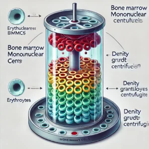
The isolation of BMMCs involves density gradient centrifugation using reagents such as Ficoll-Paque. This technique separates mononuclear cells from erythrocytes and granulocytes based on their density. Protocols for BMMC isolation can be found on Centers for Disease Control and Prevention (CDC) (cdc.gov) and U.S. Food and Drug Administration (FDA) (fda.gov).

Cryopreservation of BMMCs is achieved using dimethyl sulfoxide (DMSO) and fetal bovine serum (FBS) as cryoprotectants. Guidelines for cryopreservation are available from National Cancer Institute (NCI) (cancer.gov) and Stem Cell Research Centers at Harvard University (hscrb.harvard.edu).

Applications of BMMCs in Research and Medicine

1. Regenerative Medicine and Stem Cell Therapy

BMMCs are widely used in regenerative medicine due to their ability to differentiate into various cell types. Clinical trials using BMMCs for cardiac repair and bone regeneration are documented by ClinicalTrials.gov (clinicaltrials.gov).

BMMCs contribute significantly to research on tissue engineering, where they are used in biomaterial scaffolds to enhance tissue regeneration. The Wake Forest Institute for Regenerative Medicine (wakehealth.edu) provides research insights on this application.

2. Hematopoietic Stem Cell Transplantation (HSCT)

Hematopoietic stem cell transplantation (HSCT) using BMMCs is a life-saving therapy for leukemia, lymphoma, and other hematological diseases. For clinical guidelines, visit National Marrow Donor Program (Be The Match) (bethematch.org).

Studies also show the potential of umbilical cord blood transplantation combined with BMMCs for improved hematopoietic recovery. Research from the Fred Hutchinson Cancer Research Center (fredhutch.org) highlights the effectiveness of BMMCs in combination therapies.

3. Immunotherapy and Cancer Research

BMMCs contain immune cells such as natural killer (NK) cells and T cells, which are instrumental in immunotherapy approaches for cancer treatment. Research on immunotherapy strategies utilizing BMMCs can be found at MD Anderson Cancer Center (mdanderson.org) and National Cancer Institute (NCI) (cancer.gov).

BMMCs are also being investigated for their role in chimeric antigen receptor (CAR) T-cell therapy, which has revolutionized cancer treatment. Research insights are provided by Memorial Sloan Kettering Cancer Center (mskcc.org).

4. Autoimmune Disease Treatment

Studies have demonstrated the role of BMMCs in modulating immune responses, making them a potential therapy for autoimmune disorders such as multiple sclerosis (MS) and rheumatoid arthritis (RA). Clinical research data are available at National Institute of Arthritis and Musculoskeletal and Skin Diseases (NIAMS) (niams.nih.gov) and Johns Hopkins Autoimmune Disease Research Center (hopkinsmedicine.org).

5. Neurological Disorders and CNS Repair

Recent studies indicate that BMMCs can aid in the repair of central nervous system (CNS) damage caused by stroke, spinal cord injury, and neurodegenerative diseases like Parkinson’s and Alzheimer’s. The National Institute of Neurological Disorders and Stroke (NINDS) (ninds.nih.gov) provides further research on the subject.

Challenges and Future Directions

Despite their vast potential, the use of BMMCs presents challenges, such as donor variability, risk of contamination, and ethical considerations. Regulatory guidelines for the use of human-derived cells are provided by the U.S. Food and Drug Administration (FDA) (fda.gov) and World Health Organization (WHO) (who.int).

Ongoing research focuses on enhancing the differentiation potential of BMMCs and improving cryopreservation techniques to ensure higher post-thaw viability. Scientists at Stanford University Stem Cell Institute (med.stanford.edu) and University of California, San Francisco (UCSF) Stem Cell Program (stemcell.ucsf.edu) are actively exploring these advancements.

Conclusion

Human Bone Marrow Mononuclear Cells (BMMCs) represent a critical component in modern biomedical research and clinical applications. Their use in stem cell therapy, immunotherapy, regenerative medicine, and hematological treatments underscores their importance in advancing medical science.

For further resources, visit the National Heart, Lung, and Blood Institute (NHLBI) (nhlbi.nih.gov) and Mayo Clinic Research (mayoclinic.org). As research continues to progress, BMMCs will likely play an increasingly pivotal role in treating and understanding various diseases.